


HAWK’s data model is designed to accommodate wildlife, arthropod, and environmental health data from various sources, including local to national wildlife health surveillance systems, research initiatives, citizen-science based projects, or any other source of wildlife health information. We strongly recommend first learning about the core components of the data model: Project to Results, and Interpretation.
HAWK structures surveillance data following a hierarchy, capturing relevant epidemiological information at each step. The full hierarchy includes:
The main relationships among the basic units of the data model are shown in Figure 1 below:
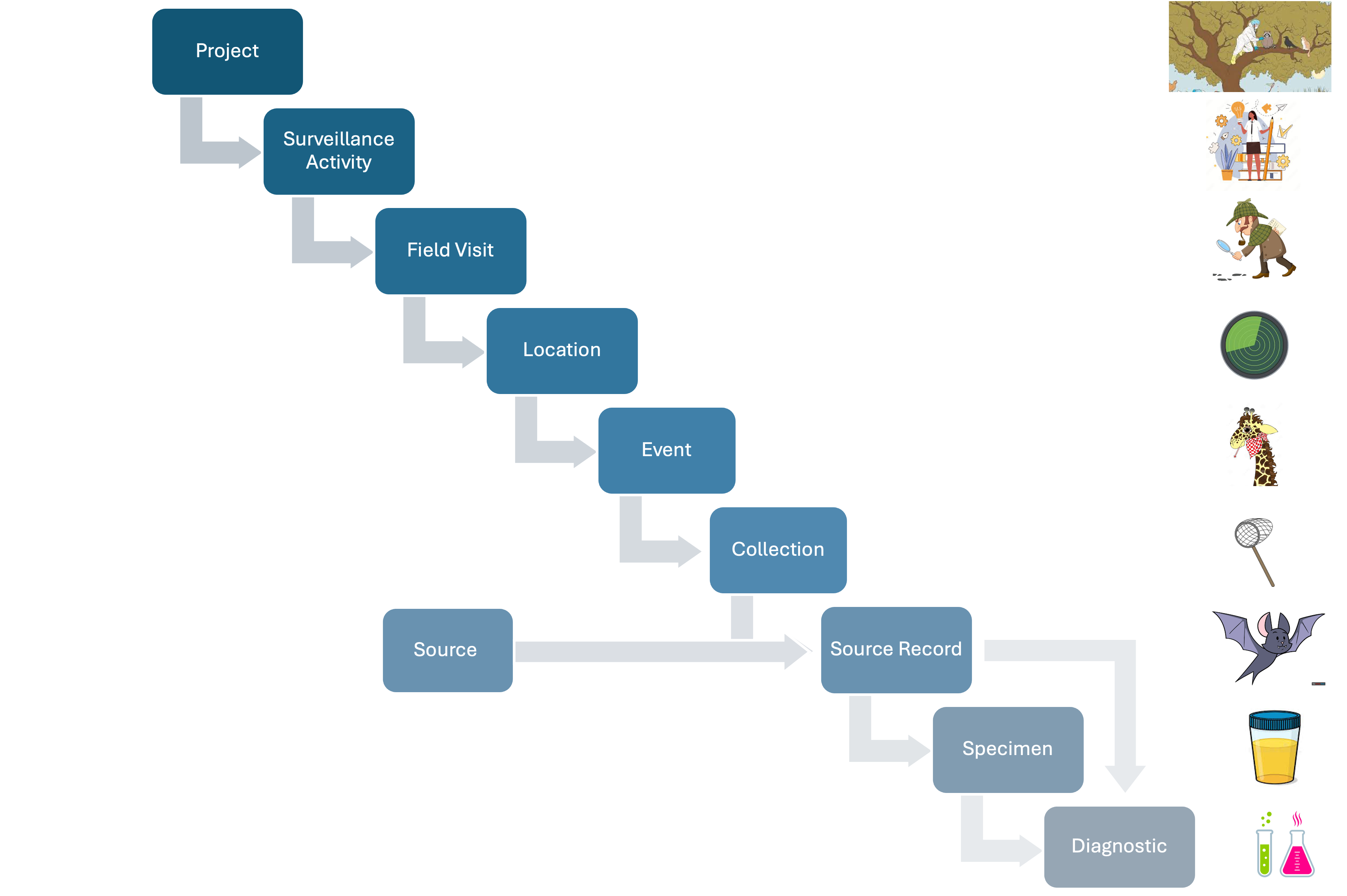
Figure 1: Basic relationships among the basic units of the data model.
HAWK employs a modular approach to the data model, enabling components to be added based on specific user needs.
When data comes from wildlife mortality reported by a community member through a mobile application:
The Surveillance Activity contains a description of the methodology
The Field Visit could be a day when the mortality was found
The Location could be the site where the mortality was found
An Event is the epidemiological unit with latitude, longitude, and time representing the position where dead animals were found
The Animal Source Records describe the dead animals
No Specimens, Diagnostics, or Results data are generated
No Effort is involved as the animals are found opportunistically
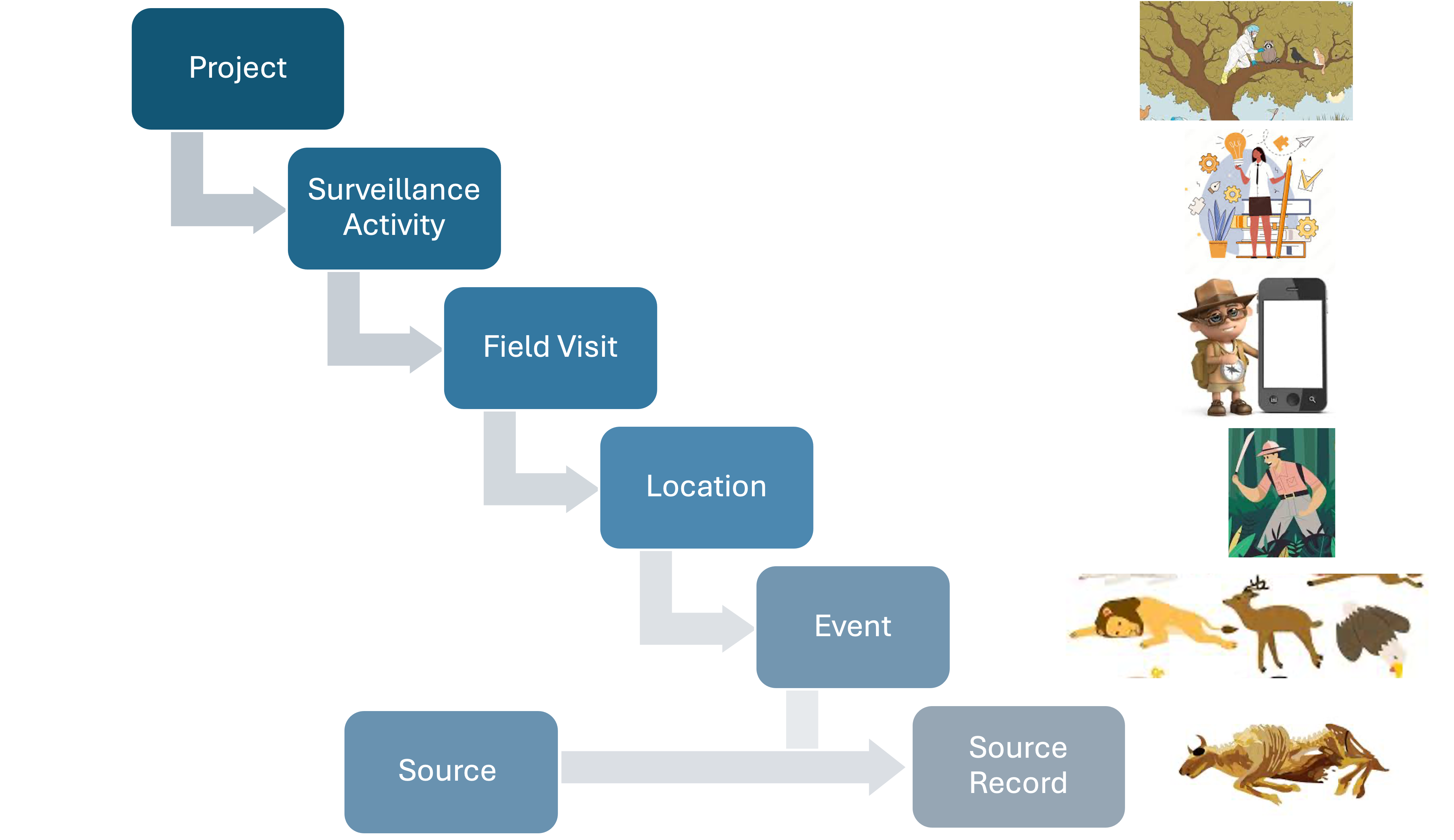
Figure Case Study 1: An illustration of how case study 1 data is structured in the data model.
This flexible structure means the model remains efficient and scalable, adapting to different users and surveillance needs.
A Project in the data model represents a planned set surveillance operations with specific goals or a common topic, usually with a defined scope and set of tasks with a common leader and organizer (e.g., a national wildlife health surveillance system) supported by specific entities and lead by specific individuals.
Projects serve as the first hierarchical unit in the database under an Organization and must contain at least one Surveillance Activity. They can be time-limited, spanning a single date, or ongoing for an extended period as needed.
Key properties of a Project include:
Other attributes are provided in the Data Dictionary (under construction)
Surveillance of New World Screwworm. The Project includes surveillance of adult flies trapped with baited devices, news sources reporting infestation cases in wildlife, and camera trap videos with wildlife showing wounds compatible with New World Screwworm infestation.
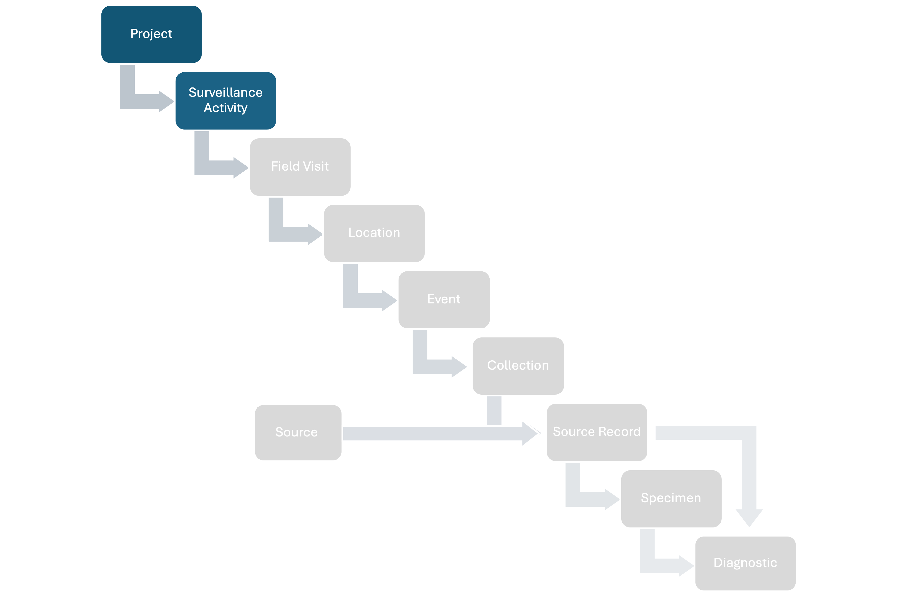 Figure 2: Postition of Surveillance Activity in the data model.
A Surveillance Activity in the data model refers to a coordinated set of activities aimed at detecting diseases, demonstrating disease freedom, measuring incidence or prevalence, or assessing trends in specific health metrics within defined populations. In general, a Surveillance Activity will be characterized by a specific objective, and corresponding methods.
Each Surveillance Activity includes metadata describing objectives and methods. Key properties of a Surveillance Activity include:
Other attributes are provided in the Data Dictionary (under construction)
A Surveillance Activity can span multiple Field Visits, Locations, and Source types (e.g., Groups, Animals, Environmental and Arthropod Sources).
The longitudinal assessment of coronavirus shedding in two Eidolon helvum bat roosts in Africa aimed understanding the patterns of coronavirus shedding through their reproductive cycle assessing common patterns in two roost separated by thousands of kilometers where the reproductive season do not match over time. The methods involved collecting a fixed number of fecal samples from the two bat roosts of the same species every month for 12 months, with subsequent testing for Coronaviridae sp. The Project in this case was USAID PREDICT and this study was a specific Surveillance Activity. Field Visits were each visit monthly to the roosts, the Locations were the two roosts, the Source Records were the bats at the sampling date each month, and the Specimens are the fecal samples taken from the roost
Each outbreak investigation is considered a Surveillance Activity. An Outbreak Surveillance Activity can encompass the initial Field Visit that led to the outbreak detection and subsequent Field Visits associated with the investigation and control. The initial Field Visit that led to the outbreak detection may have already been part of another Surveillance Activity and also the Outbreak Surveillance Activity (e.g., routine monitoring of a particular area), and this relationship is maintained to allow traceability on the history of detections.
For instance, an outbreak may first be detected during routine patrols by rangers. In such cases, the initial ranger-conducted Field Visit and its components should belong to both the ranger patrol Surveillance Activity and also the outbreak Surveillance Activity. Follow-up Field Visits (e.g., by veterinarians or public health officers investigating the outbreak) should be assigned exclusively to the outbreak Surveillance Activity.
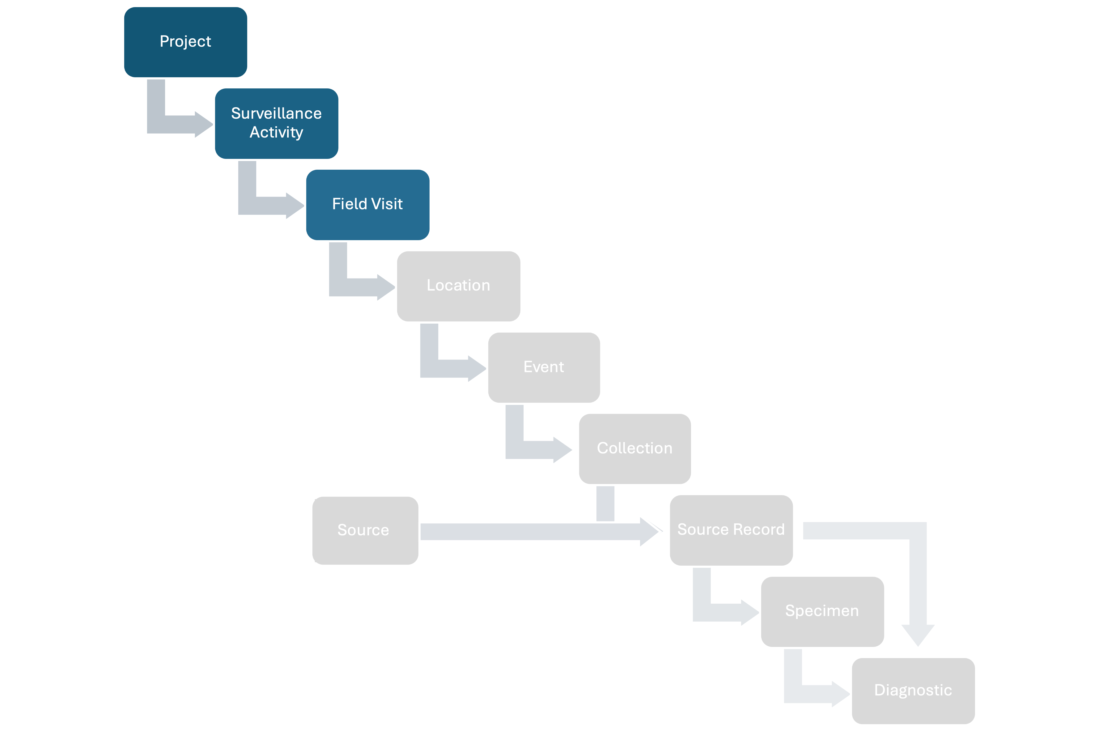 Figure 3: Position of Field Visit in the data model.
A Field Visit in the data model represents a defined time period—including a start and end date—during which Locations are visited, Events and Sources are conducted, identified, or observed, and Specimens might be collected and documented. The length of a Field Visit is flexible. The meaning of a Field Visit depends on the methodology of the Surveillance Activity. Users must define what a Field Visit represents and document it in the Surveillance Activity metadata.
Key properties of a Field Visit include:
Other attributes are provided in the Data Dictionary (under construction)
In the monitoring of wildlife anomalies through camera traps (for example, as described here), a Field Visit refers to the period during which a set of camera traps was deployed in the field
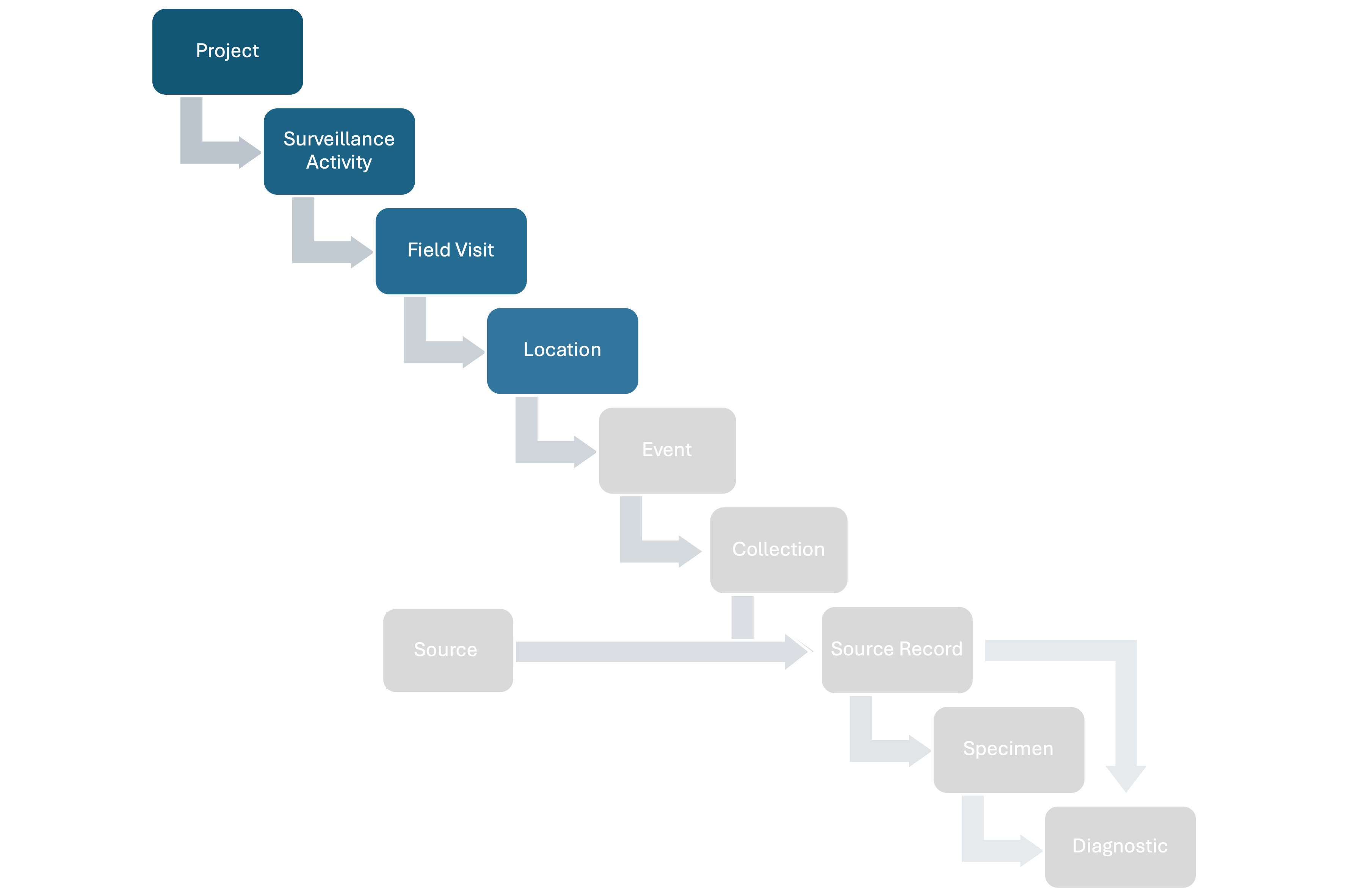 Figure 4: Position of Location in the data model.
In the data model, a Location represents a general area where Field Visits take place. Locations are not defined by exact latitude and longitude coordinates (unlike Events, defined later), but they serve as a broader spatial grouping for organizing data. The meaning of a Location depends on the methodology of the Surveillance Activity. Users must define what a Location represents and document it in the Surveillance Activity metadata.
Key attributes of a Location include:
Other attributes are provided in the Data Dictionary (under construction)
In the sampling of wildlife in markets, a Location can refer to a specific market
In the data model, Effort contains the information associated with the spatial and temporal metrics used to activel obtain Sources in the field.
When Source Records are not linked to an Effort (e.g., opportunist finding of dead animals), then no Effort is involved.
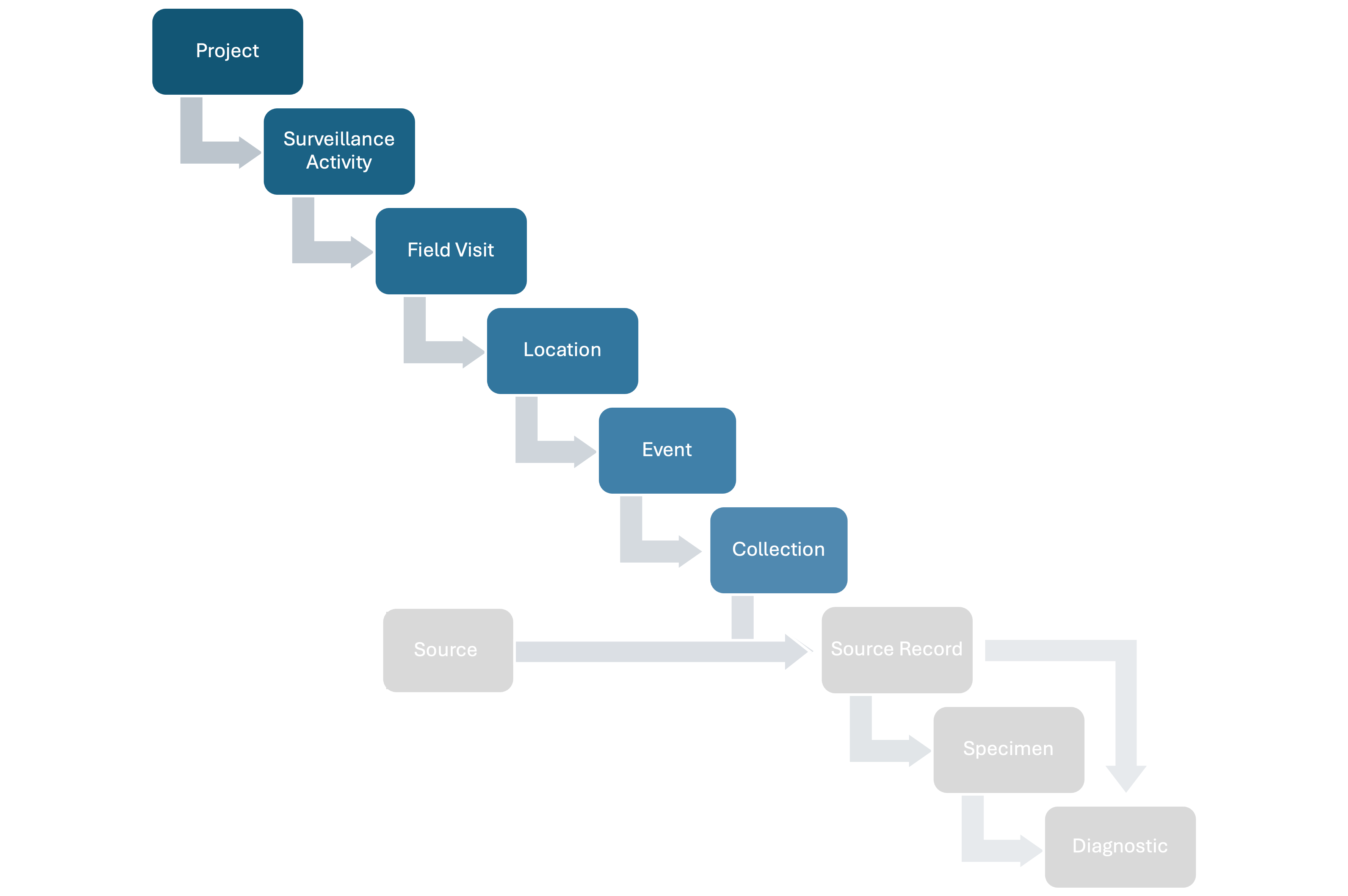 Figure 5: Position of Effort in the data model.
Area Effort refers to the spatial and temporal work to find or document Events within an area (e.g., square kilometers explored in x amount of time) within a Location. The meaning of Area Effort depends on the methodology of the Surveillance Activity. Users must define what a Area Effort represents and document it in the Surveillance Activity metadata.
Key attributes of a Area Effort include:
Other attributes are provided in the Data Dictionary (under construction)
Following recommendations here, in an outbreak investigation, dead marine birds are actively searched in a beach. In this case, the beach is the Location and each area explored within the beach during a specific period of time represents an Area Effort
A Track Effort refers to the spatial and temporal work to find or document Events in linear features (e.g., kilometers walked through a set of spatial coordinates in an x period of time) within a Location. The meaning of a Track Effort depends on the methodology of the Surveillance Activity. Users must define what a Track Effort represents and document it in the Surveillance Activity metadata.
Key attributes of a Track Effort include:
Other attributes are provided in the Data Dictionary (under construction)
Following the ranger-based wildlife morbidity and mortality surveillance described here, a sector within a protected area is the Location and the route traveled by patrolling rangers to find wildlife morbidity and mortality during a specific period of time represents a Track Effort.
Track Efforts are not nested in Areas Efforts. If a methodology involves Events nested in Track Efforts nested in Area Efforts then, the link between both types of Effort can be made through the spatial data and through Tags (see “Advanced”).
A Point Effort refers to the spatial and temporal work to find or document Events at a specific spatio-temporal coordinate (e.g., a device set up at a specific longitude and latitud for x amount of time to capture mosquitoes) within a Location. The meaning of a Point Effort depends on the methodology of the Surveillance Activity. Users must define what a Point Effort represents and document it in the Surveillance Activity metadata.
Key attributes of a Point Effort include:
Other attributes are provided in the Data Dictionary (under construction)
In the counting of dead birds at a wetland from a tower and using binoculars, the Point Collection is the position and number of observers recording dead birds from the tower during a specific period of time
In the monitoring of wildlife morbidity through camera traps, the Point Collection is the number camera traps deployed at a specific position to photograph animals and the hours or days active.
Area, Track and Point Effort fields also help capture issues encountered when searching for Sources, such as camera traps running out of power, being stolen, or damaged mist nets.
Efforts can contain between zero (e.g., an unsuccessful capture where no animals are caught) and an unlimited number of Source Records**.
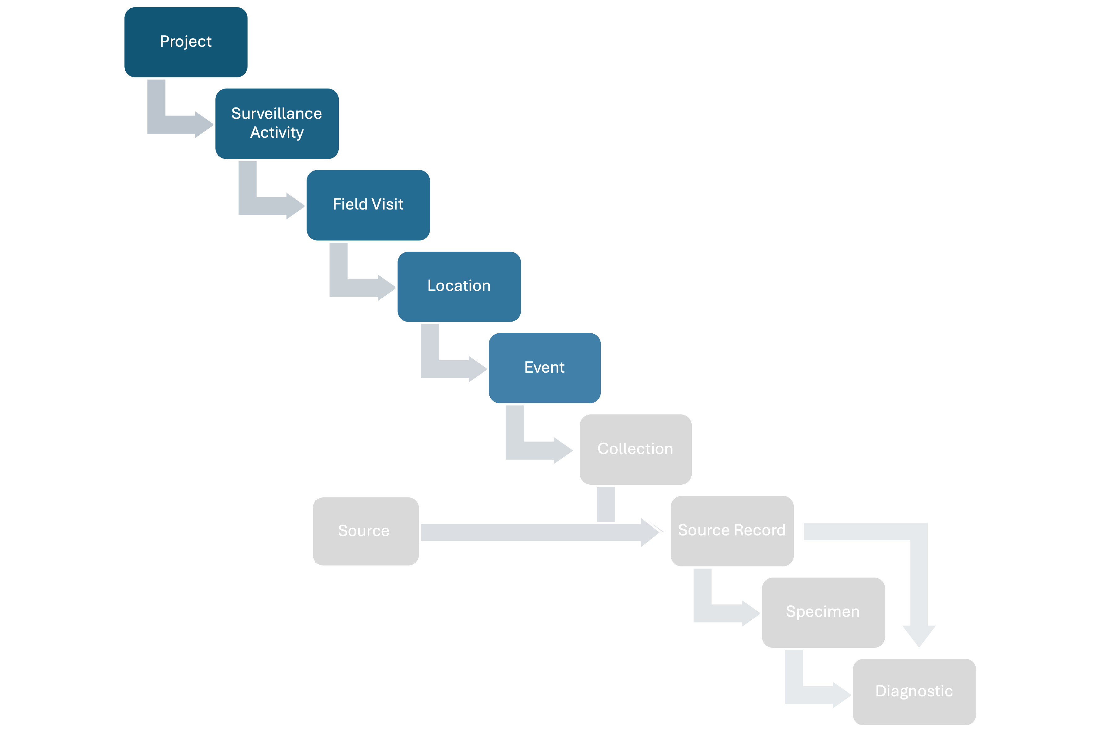 Figure 6: Position of Event in the data model.
In the data model, an Event represents a distinct spatio-temporal coordinate where Source Record data is collected from (e.g., specific longitude, latitude, and timestamp). The definition of Event depends on the methodology of the Surveillance Activity. Users must define what an Event represents and document it in the Surveillance Activity metadata. We strongly recommend that an Event represents the minimal epidemiological unit of interest containing Source Records at position p and time t (e.g., the sampling event of animals in a cage, the collection of mosquitoes using a trap, the position of a dead animal found in the field at time t)
Key attributes of a Event include:
Other attributes are provided in the Data Dictionary (under construction)
A pair of mist nets are deployed to capture bats and sample them. The Event in this case is the sampling of the bats captured at time t using the mist nets. The nets are part of a Point Effort. In this case, the Event receives the same spatial coordinates as the Point Effort
A dead animal is found during a ranger patrol. In this case the Event is the position of the dead animal and the time of encounter
Injured animals are recorded using a camera trap. In this case the Event is each capture of animals by the camera trap and they receive the same spatial coordinates as the Point Effort. The Point Effort is the camera traps at a specific spatial position and the deployment time
For more examples see here
Other relevant characteristics of an Event include:
An Event might not belong to a Area, Track, or Point Effort. For example, the position of dead birds accidentally found in a beach or sampling of the animals in cages in a wet market.
An Event can include more than one Source type (see next section for a full description of Sources). For example, mosquito traps and collection of wastewater at the same position p and time t.
An Event can include or not at-observation normal (“healthy”) animals depending on the definition of Event.
An Event might not contain any Source Records (see next section for a full description of Sources). For example, when a mist net is deployed but it fails to capture any animal. The mist net (Point Effort is deployed, the sampling Event is conducted at time t but no animal is captured).
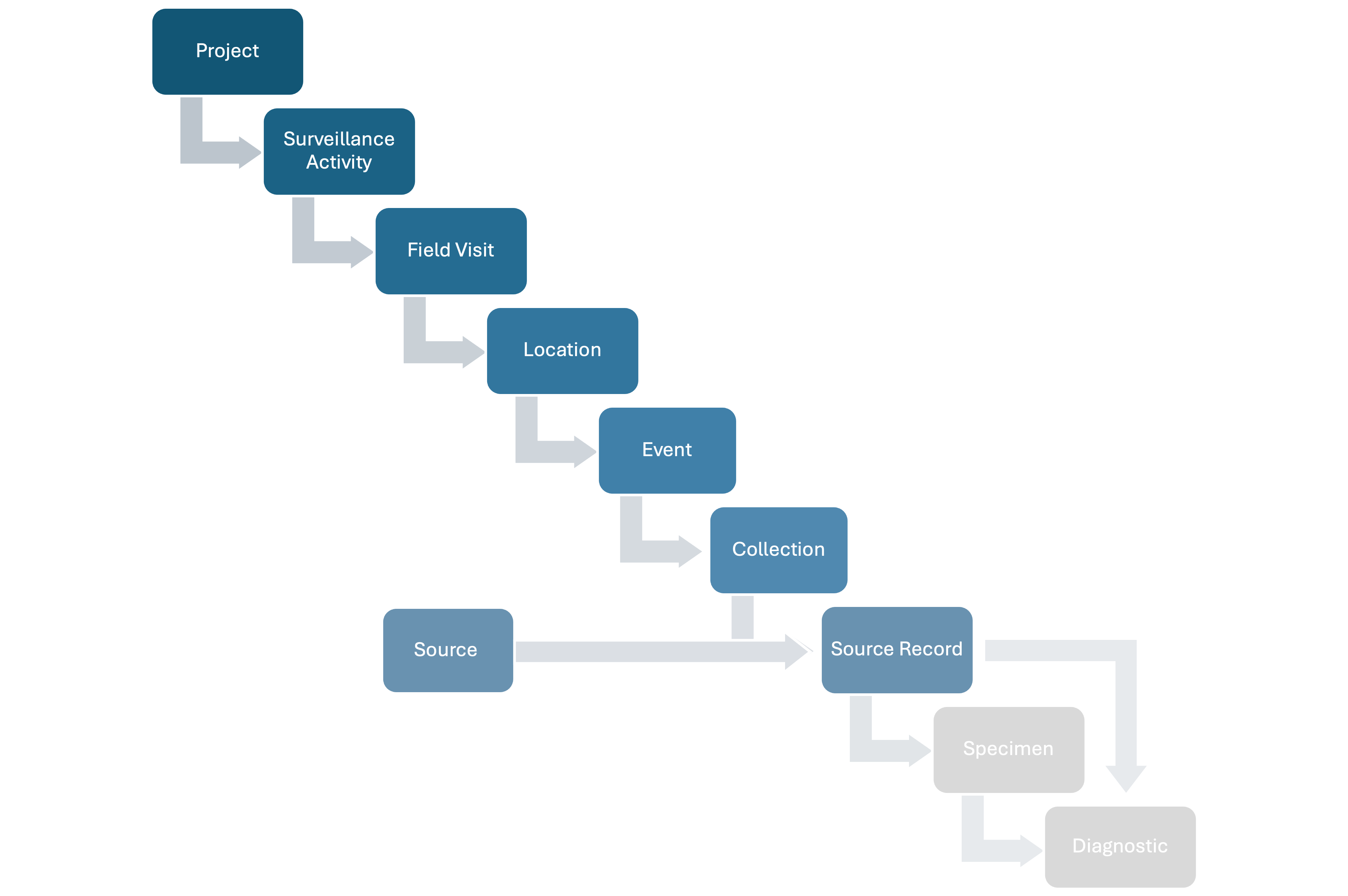 Figure 8: Position of Source Records in the data model.
In the data model, a Source is an epidemiological unit of interest that can be observed, necropsied, diagnosed, or provide Specimens for analysis. The data model manages four types of Sources: i) a group of individuals of the same taxa (Group Source), ii) individual animal (Animal Source), iii) a site providing arthropods (Arthropod Source), and iv) a site providing environmental abiotic specimens (e.g., wastewater) or biotic specimens (e.g., feces not linked to a specific animal) or where metrics such are pH can be measured at (Environmental Source).
Identified Sources can be found at position p and time t in a single or multiple Events.
A Group Source is a unit of individuals of the same taxa that belong to a common entity, such as a herd, roost, area, site, farm, cage, stall, or enclosure, forming a single epidemiological unit. Group Sources can be observed, captured, diagnosed, and interpreted as a full unit, and provide collective Specimens if sampling is conducted. A Group Source can be shipped and stored when the individuals are dead.
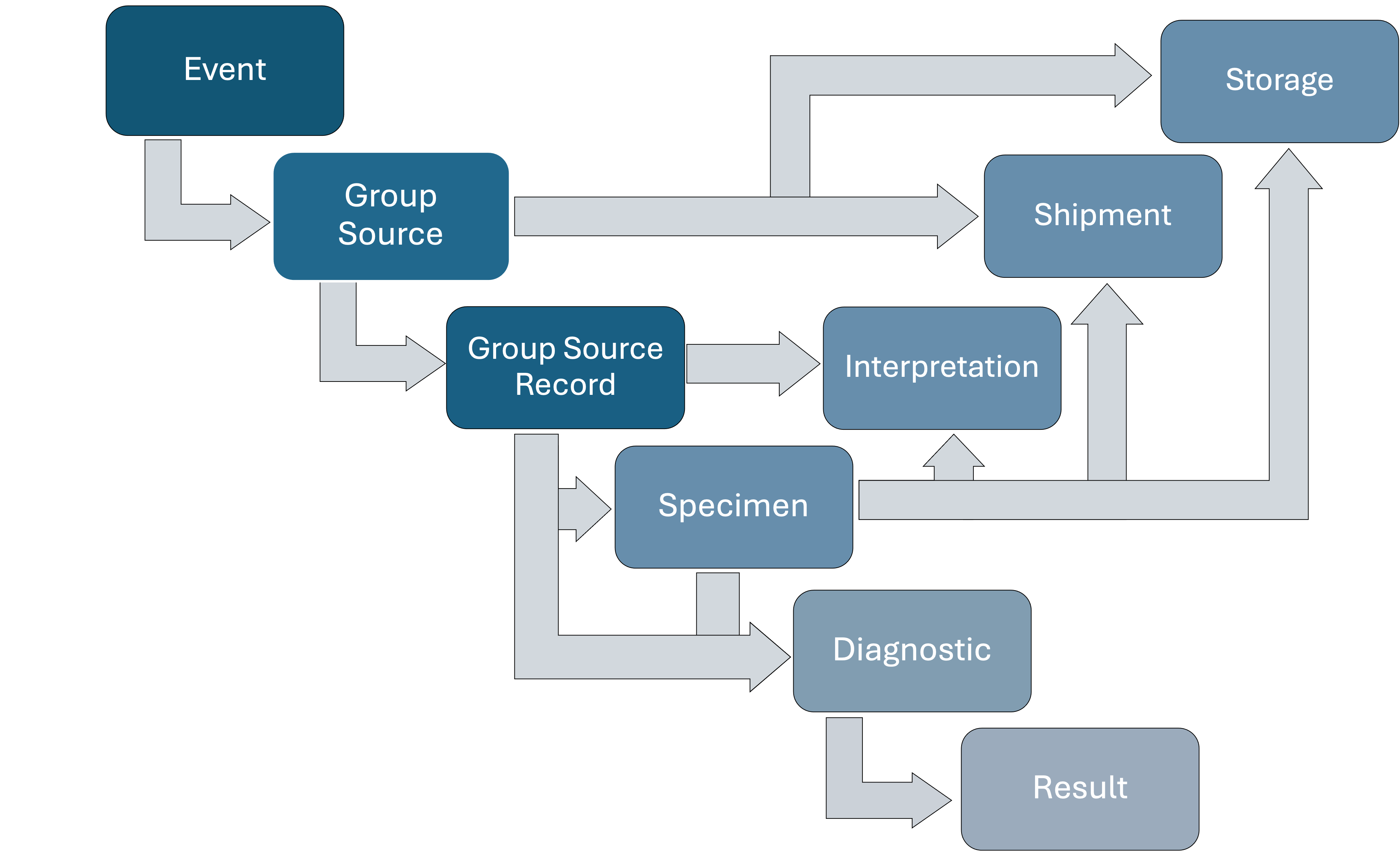 Figure 9: Detailed relationships of Group Source in the data model.
A Group Source Record documents the animals of a Group Source at time t, categorized by sex, age, and life status (e.g., live or dead).
Key attributes of a Group Source Record include time independent variables:
Other attributes are provided in the Data Dictionary (under construction)
Several attributes are recorded at the group level and, therefore, multiple options can be included. For example, several observed anomalies may be listed for a Group Source Record, but their distribution across individuals cannot be recorded.
A Group Source Record can be used for a Diagnostic (e.g., assessing the body condition of a specific herd using a standardized metric).
Carcasses of a Group Source Record can be recorded collectively (e.g., multiple dead frogs in a plastic bag) and they can be stored, shipped, and used collectively in a diagnostic test (see “Storage”, “Shipments”, and “Diagnostic” below).
Carcasses of a Group Source Record cannot have a collective necropsy. The individuals animals must be converted to Animal Sources.
The total number of animals of a specific taxon in an Event is the sum of the individuals of the taxa in the Group Source Record and each individual animal of the same taxa recorded as Animal Source Record. For example, consider a herd of 20 cows illegally raised in a protected area whose health is assessed at time t. If two cows are sampled, the two sampled cows should be documented as two separate Animal Source Records (one per cow), while the remaining 18 cows are recorded as a single Group Source Record, categorized by sex, age, and life status. In this scenario, the total number of cows is 20—the 18 recorded in the Group Source Record plus the two documented as Animal Source Records. The “herd” identity of these 20 cows split in three Source Records can be maintained using tags (see Complexities). If, however, the Group Source Record incorrectly includes all 20 cows while two of them are also recorded as Animal Source Record, the total count would incorrectly sum to 22.
An Animal Source represents a single individual. Animal Sources can be observed, captured, tested, necropsied, interpreted, and provide Specimens directly or during a Necropsy. An Animal Source can be shipped and stored if the individual is dead.
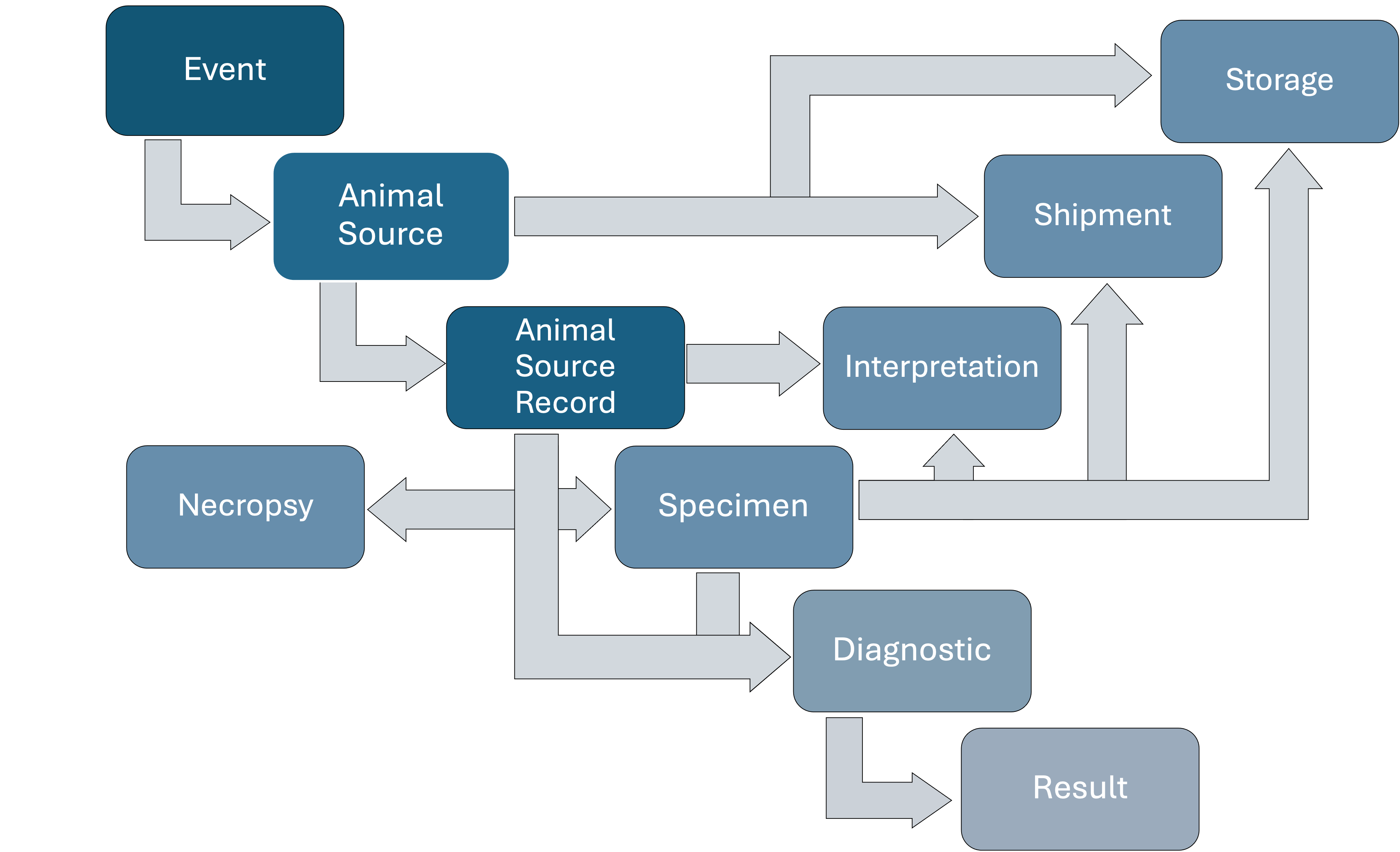 Figure 10: Detailed relationships of Animal Source in the data model.
An Animal Source Record represents an individual Animal Source at time t.
Key attributes of a Animal Source Record include:
Other attributes are provided in the Data Dictionary (under construction)
Some properties of an Animal Source Record allow single or multiple selections. For instance, an animal’s life status is recorded as one category (e.g., “live” or “dead”), whereas multiple anomalies may be reported for the same individual at time t (e.g., wounds, hair loss, diarrhea).
An Animal Source Record can be categorized as dead or live at time t. A live animal at time t can be part of an Event, depending on the Event’s definition.
An Animal Source Record can be used directly for a Diagnostic (e.g., performing X-rays on an animal).
Vaccinations administered to an Animal Source Record can be added ensuring that vaccination history accumulates.
The carcass of an Animal Source Record can be stored, shipped, and used in a diagnostic test (see “Storage”, “Shipments”, and “Diagnostic” below). Live Animal Sources cannot can be shipped or stored.
The carcass of an Animal Source Record can be used in a Necropsy (see “Necropsy” below).
The total number of animals of a specific taxon in an Event is the sum of the individuals of the taxa in the Group Source Record and each individual animal of the same taxa recorded as Animal Source Record. For example, consider a herd of 20 cows illegally raised in a protected area whose health is assessed at time t. If two cows are sampled, the two sampled cows should be documented as two separate Animal Source Records (one per cow), while the remaining 18 cows are recorded as a single Group Source Record, categorized by sex, age, and health status. In this scenario, the total number of cows is 20—the 18 recorded in the Group Source Record plus the two documented as Animal Source Records. The “herd” identity of these 20 cows split in three Source Records can be maintained using a cluster (see Complexities). If, however, the Group Source Record incorrectly includes all 20 cows while two of them are also recorded as Animal Source Record, the total count would incorrectly sum to 22.
A Necropsy is linked to a specific Animal Source Record. A Necropsy can take place in the field or in a facility.
Key attributes of a Necropsy include:
Other attributes are provided in the Data Dictionary (under construction)
The Necropsy findings are reported through text in fields such as “Gastrointestinal System”, “Reproductive System”, etc. The list of anatomical components encompassed by each of these elements or systems is provided in the table below and they follow the National Agricultural Library Thesaurus Concept Space (NALT), a controlled vocabulary of terms related to agricultural, biological, physical and social sciences (https://lod.nal.usda.gov/nalt/en/).
| Topography | NALT_URI | Definition_NALT | Alternative_name |
|---|---|---|---|
| retina | https://lod.nal.usda.gov/nalt/38995 | The ten-layered nervous tissue membrane of the eye. It is continuous with the optic nerve and receives images of external objects and transmits visual impulses to the brain. Its outer surface is in contact with the choroid and the inner surface with the vitreous body. The outer-most layer is pigmented, whereas the inner nine layers are transparent. | NA |
| eye lens | https://lod.nal.usda.gov/nalt/38989 | NA | |
| sclera | https://lod.nal.usda.gov/nalt/290574 | The white, opaque, fibrous, outer tunic of the eyeball, covering it entirely excepting the segment covered anteriorly by the cornea. It is essentially avascular but contains apertures for vessels, lymphatics, and nerves. It receives the tendons of insertion of the extraocular muscles and at the corneoscleral junction contains the canal of Schlemm. | eye white |
| eyes | https://lod.nal.usda.gov/nalt/28392 | eye | |
| lacrimal apparatus | https://lod.nal.usda.gov/nalt/28564 | lacrimal gland | |
| lacrimal apparatus | https://lod.nal.usda.gov/nalt/28564 | conjunctival sac | |
| iris (eyes) | https://lod.nal.usda.gov/nalt/38993 | The most anterior portion of the uveal layer, separating the anterior chamber from the posterior. It consists of two layers - the stroma and the pigmented epithelium. Color of the iris depends on the amount of melanin in the stroma on reflection from the pigmented epithelium. | NA |
| conjunctiva | https://lod.nal.usda.gov/nalt/28561 | NA | |
| cornea | https://lod.nal.usda.gov/nalt/29206 | NA | |
| eyelids | https://lod.nal.usda.gov/nalt/38991 | NA |
| Topography | NALT_URI | Definition_NALT | Alternative_name |
|---|---|---|---|
| inner ear | https://lod.nal.usda.gov/nalt/332326 | The essential part of the hearing organ consists of two labyrinthine compartments: the bony labyrinthine and the membranous labyrinth. The bony labyrinth is a complex of three interconnecting cavities or spaces (Cochlea; vestibular labyrinth; and semicircular canals) in the temporal bone. Within the bony labyrinth lies the membranous labyrinth which is a complex of sacs and tubules (cochlear duct; saccule and utricle; and semicircular ducts) forming a continuous space enclosed by epithelium and connective tissue. These spaces are filled with labyrinthine fluids of various compositions. | labyrinth (inner ear) |
| guttural pouch | https://lod.nal.usda.gov/nalt/35107 | NA | |
| ears | https://lod.nal.usda.gov/nalt/35074 | ear | |
| middle ear | https://lod.nal.usda.gov/nalt/332330 | The space and structures directly internal to the tympanic membrane and external to the inner ear (labyrinth). Its major components include the auditory ossicles and the eustachian tube that connects the cavity of middle ear (tympanic cavity) to the upper part of the throat. | tympanum |
| middle ear | https://lod.nal.usda.gov/nalt/332330 | The space and structures directly internal to the tympanic membrane and external to the inner ear (labyrinth). Its major components include the auditory ossicles and the eustachian tube that connects the cavity of middle ear (tympanic cavity) to the upper part of the throat. | tympanic cavity |
| otoliths | https://lod.nal.usda.gov/nalt/191711 | Structures of the inner ear which function in orientation and equilibrium sensing of vertebrates. Otoliths contain calcium concretions useful for species identification and age determination of fish, and provide information about their life history and ecology. | otoconia |
| otoliths | https://lod.nal.usda.gov/nalt/191711 | Structures of the inner ear which function in orientation and equilibrium sensing of vertebrates. Otoliths contain calcium concretions useful for species identification and age determination of fish, and provide information about their life history and ecology. | otolithic membrane |
| otoliths | https://lod.nal.usda.gov/nalt/191711 | Structures of the inner ear which function in orientation and equilibrium sensing of vertebrates. Otoliths contain calcium concretions useful for species identification and age determination of fish, and provide information about their life history and ecology. | statoconia |
| otoliths | https://lod.nal.usda.gov/nalt/191711 | Structures of the inner ear which function in orientation and equilibrium sensing of vertebrates. Otoliths contain calcium concretions useful for species identification and age determination of fish, and provide information about their life history and ecology. | otolites |
| ear canal | https://lod.nal.usda.gov/nalt/339897 | The narrow passage way that conducts the sound collected by the ear auricle to the tympanic membrane. | auditory canal |
| ear canal | https://lod.nal.usda.gov/nalt/339897 | The narrow passage way that conducts the sound collected by the ear auricle to the tympanic membrane. | meatus acusticus externus |
| Topography | NALT_URI | Definition_NALT | Alternative_name |
|---|---|---|---|
| nose | https://lod.nal.usda.gov/nalt/54351 | nostrils | |
| vomeronasal organ | https://lod.nal.usda.gov/nalt/308291 | An accessory chemoreceptor organ that is separated from the main olfactory mucosa. It is situated at the base of nasal septum close to the vomer and nasal bones. It forwards chemical signals (such as pheromones) to the central nervous system, thus influencing reproductive and social behavior. In humans, most of its structures except the vomeronasal duct undergo regression after birth. | Jacobson organ |
| vomeronasal organ | https://lod.nal.usda.gov/nalt/308291 | An accessory chemoreceptor organ that is separated from the main olfactory mucosa. It is situated at the base of nasal septum close to the vomer and nasal bones. It forwards chemical signals (such as pheromones) to the central nervous system, thus influencing reproductive and social behavior. In humans, most of its structures except the vomeronasal duct undergo regression after birth. | Jacobson's organ |
| vomeronasal organ | https://lod.nal.usda.gov/nalt/308291 | An accessory chemoreceptor organ that is separated from the main olfactory mucosa. It is situated at the base of nasal septum close to the vomer and nasal bones. It forwards chemical signals (such as pheromones) to the central nervous system, thus influencing reproductive and social behavior. In humans, most of its structures except the vomeronasal duct undergo regression after birth. | vomeronasal nerve |
| nasal mucosa | https://lod.nal.usda.gov/nalt/53591 | NA | |
| nasal cavity | https://lod.nal.usda.gov/nalt/54350 | NA |
| Topography | NALT_URI | Definition_NALT | Alternative_name |
|---|---|---|---|
| teeth | https://lod.nal.usda.gov/nalt/32369 | dentine | |
| teeth | https://lod.nal.usda.gov/nalt/32369 | dentition | |
| beak | https://lod.nal.usda.gov/nalt/16134 | NA | |
| gingiva | https://lod.nal.usda.gov/nalt/9531 | Oral tissue surrounding and attached to teeth. | interdental papilla |
| gingiva | https://lod.nal.usda.gov/nalt/9531 | Oral tissue surrounding and attached to teeth. | gums (mouth) |
| lips | https://lod.nal.usda.gov/nalt/50308 | NA | |
| salivary glands | https://lod.nal.usda.gov/nalt/2899 | NA | |
| palate | https://lod.nal.usda.gov/nalt/26789 | roof of the mouth | |
| palate | https://lod.nal.usda.gov/nalt/26789 | hard palate | |
| palate | https://lod.nal.usda.gov/nalt/26789 | soft palate | |
| mouth | https://lod.nal.usda.gov/nalt/33560 | buccal cavity | |
| mouth | https://lod.nal.usda.gov/nalt/33560 | oral cavity | |
| mouth | https://lod.nal.usda.gov/nalt/33560 | periodontium | |
| tongue | https://lod.nal.usda.gov/nalt/53559 | NA |
| Topography | NALT_URI | Definition_NALT | Alternative_name |
|---|---|---|---|
| Insect cuticle | https://lod.nal.usda.gov/nalt/9104 | NA | NA |
| Perineum | https://lod.nal.usda.gov/nalt/57466 | NA | NA |
| Scar | NA | NA | NA |
| Scent glands | https://lod.nal.usda.gov/nalt/59050 | NA | Poll glands |
| Sebaceous glands | https://lod.nal.usda.gov/nalt/38913 | NA | Tarsal glands |
| Skin (animal) | https://lod.nal.usda.gov/nalt/32499 | NA | NA |
| Sweat glands | https://lod.nal.usda.gov/nalt/38914 | NA | NA |
| Wounds and injuries | https://lod.nal.usda.gov/nalt/728 | NA | NA |
| Antlers | https://lod.nal.usda.gov/nalt/10565 | NA | NA |
| Claws | https://lod.nal.usda.gov/nalt/26735 | NA | Talons |
| Claws | https://lod.nal.usda.gov/nalt/26735 | NA | Nails (non-primates) |
| Claws | https://lod.nal.usda.gov/nalt/26735 | NA | Claw horn |
| Comb (integument) | https://lod.nal.usda.gov/nalt/28099 | NA | NA |
| Eyelashes | NA | NA | NA |
| Feathers | https://lod.nal.usda.gov/nalt/34442 | NA | NA |
| Fur | https://lod.nal.usda.gov/nalt/27376 | NA | Coat (animal) |
| Hairs | https://lod.nal.usda.gov/nalt/44301 | NA | Scalp hair |
| Hairs | https://lod.nal.usda.gov/nalt/44301 | NA | Hairs (animal) |
| Hooves | https://lod.nal.usda.gov/nalt/45820 | NA | Hoofs |
| Integument | https://lod.nal.usda.gov/nalt/9103 | NA | Exuviae |
| Integument | https://lod.nal.usda.gov/nalt/9103 | NA | Body wall |
| Nails (integument) | https://lod.nal.usda.gov/nalt/47469 | NA | Nails (primates) |
| Nails (integument) | https://lod.nal.usda.gov/nalt/47469 | NA | Nails (human) |
| Scales (integument) | https://lod.nal.usda.gov/nalt/47470 | NA | NA |
| Wattles | https://lod.nal.usda.gov/nalt/47472 | NA | NA |
| Topography | NALT_URI | Definition_NALT | Alternative_name |
|---|---|---|---|
| subcutaneous fat | https://lod.nal.usda.gov/nalt/18338 | NA |
| Topography | NALT_URI | Definition_NALT | Alternative_name |
|---|---|---|---|
| joints (animal) | https://lod.nal.usda.gov/nalt/36135 | NA | |
| tendons | https://lod.nal.usda.gov/nalt/28572 | Fibrous bands or cords of connective tissue at the ends of muscle fibers that serve to attach the muscles to bones and other structures. | NA |
| bones | https://lod.nal.usda.gov/nalt/18497 | NA | |
| skeletal muscle | https://lod.nal.usda.gov/nalt/35050 | NA | |
| fascia | https://lod.nal.usda.gov/nalt/28570 | NA | |
| skeleton | https://lod.nal.usda.gov/nalt/53793 | NA |
| Topography | NALT_URI | Definition_NALT | Alternative_name |
|---|---|---|---|
| mediastinum | https://lod.nal.usda.gov/nalt/51800 | NA | |
| body cavities | https://lod.nal.usda.gov/nalt/1610 | NA | |
| abdominal cavity | https://lod.nal.usda.gov/nalt/1609 | NA | |
| hemocoel | https://lod.nal.usda.gov/nalt/18328 | A body cavity of arthropods and some molluscs that contains blood or hemolymph, which functions as part of the circulatory system by directly bathing the organs. | haemocoel |
| thoracic cavity | https://lod.nal.usda.gov/nalt/18330 | NA |
| Topography | NALT_URI | Definition_NALT | Alternative_name |
|---|---|---|---|
| Heart | https://lod.nal.usda.gov/nalt/9341 | NA | Endocardium |
| Blood plasma | https://lod.nal.usda.gov/nalt/18062 | NA | NA |
| Arteries | https://lod.nal.usda.gov/nalt/10603 | The vessels carrying blood away from the heart. | NA |
| Inferior vena cava | https://lod.nal.usda.gov/nalt/190442 | The venous trunk which receives blood from the lower extremities and from the pelvic and abdominal organs. | NA |
| Blood veins | https://lod.nal.usda.gov/nalt/18176 | NA | NA |
| Blood vessels | https://lod.nal.usda.gov/nalt/12438 | NA | NA |
| Jugular vein | https://lod.nal.usda.gov/nalt/18177 | NA | NA |
| Coronary vessels | https://lod.nal.usda.gov/nalt/12440 | The veins and arteries of the heart. | Coronary arteries |
| Coronary sinus | https://lod.nal.usda.gov/nalt/190439 | A short vein that collects about two thirds of the venous blood from the myocardium and drains into the right atrium. Coronary sinus, normally located between the left atrium and left ventricle on the posterior surface of the heart, can serve as an anatomical reference for cardiac procedures. | NA |
| Hemolymph | https://lod.nal.usda.gov/nalt/26359 | Bloodlike fluid of the hemocoel in open circulatory systems of arthropods and most molluscs. | Haemolymph |
| Portal vein | https://lod.nal.usda.gov/nalt/18178 | NA | NA |
| Blood | https://lod.nal.usda.gov/nalt/18059 | NA | NA |
| Iliac artery | https://lod.nal.usda.gov/nalt/12442 | NA | NA |
| Hepatic artery | https://lod.nal.usda.gov/nalt/12441 | NA | NA |
| Superior vena cava | https://lod.nal.usda.gov/nalt/190443 | The venous trunk which returns blood from the head, neck, upper extremities and chest. | NA |
| Blood serum | https://lod.nal.usda.gov/nalt/18139 | The clear, watery portion of blood that separates out when blood coagulates. It lacks blood clotting factors such as fibrogen and prothrombin. | Serum |
| Carotid arteries | https://lod.nal.usda.gov/nalt/12439 | NA | NA |
| Caudal vein | https://lod.nal.usda.gov/nalt/254549 | NA | Tail vein |
| Myocardium | https://lod.nal.usda.gov/nalt/22397 | The muscle tissue of the heart. It is composed of striated, involuntary muscle cells connected to form the contractile pump to generate blood flow. | Cardiac muscle |
| Aorta | https://lod.nal.usda.gov/nalt/10602 | NA | Aortic root |
| Thymocytes | https://lod.nal.usda.gov/nalt/50929 | NA | NA |
| Fetal bovine serum | https://lod.nal.usda.gov/nalt/191461 | NA | Fetal calf serum |
| Fetal bovine serum | https://lod.nal.usda.gov/nalt/191461 | NA | Fbs (fetal bovine serum) |
| Pulmonary artery | https://lod.nal.usda.gov/nalt/12444 | NA | NA |
| Umbilical arteries | https://lod.nal.usda.gov/nalt/12445 | NA | NA |
| Saphenous vein | https://lod.nal.usda.gov/nalt/18179 | The vein which drains the foot and leg. | NA |
| Ductus arteriosus | https://lod.nal.usda.gov/nalt/28513 | NA | NA |
| Umbilical veins | https://lod.nal.usda.gov/nalt/18180 | NA | NA |
| Heart atrium | https://lod.nal.usda.gov/nalt/13743 | NA | NA |
| Vena cava | https://lod.nal.usda.gov/nalt/18181 | NA | Venae cavae |
| Heart valves | https://lod.nal.usda.gov/nalt/44757 | NA | NA |
| Heart ventricle | https://lod.nal.usda.gov/nalt/44758 | NA | NA |
| Mesenteric arteries | https://lod.nal.usda.gov/nalt/12443 | NA | NA |
| Topography | NALT_URI | Definition_NALT | Alternative_name |
|---|---|---|---|
| Lungs | https://lod.nal.usda.gov/nalt/9345 | NA | NA |
| Larynx | https://lod.nal.usda.gov/nalt/49283 | NA | NA |
| Spiracles | https://lod.nal.usda.gov/nalt/61125 | NA | NA |
| Bronchi | https://lod.nal.usda.gov/nalt/20020 | The larger air passages of the lungs arising from the terminal bifurcation of the trachea. They include the largest two primary bronchi which branch out into secondary bronchi, and tertiary bronchi which extend into bronchioles and pulmonary alveoli. | Secondary bronchi |
| Bronchi | https://lod.nal.usda.gov/nalt/20020 | The larger air passages of the lungs arising from the terminal bifurcation of the trachea. They include the largest two primary bronchi which branch out into secondary bronchi, and tertiary bronchi which extend into bronchioles and pulmonary alveoli. | Primary bronchi |
| Bronchi | https://lod.nal.usda.gov/nalt/20020 | The larger air passages of the lungs arising from the terminal bifurcation of the trachea. They include the largest two primary bronchi which branch out into secondary bronchi, and tertiary bronchi which extend into bronchioles and pulmonary alveoli. | Bronchus |
| Bronchi | https://lod.nal.usda.gov/nalt/20020 | The larger air passages of the lungs arising from the terminal bifurcation of the trachea. They include the largest two primary bronchi which branch out into secondary bronchi, and tertiary bronchi which extend into bronchioles and pulmonary alveoli. | Tertiary bronchi |
| Air sacs | https://lod.nal.usda.gov/nalt/5852 | NA | NA |
| Vomeronasal organ | https://lod.nal.usda.gov/nalt/308291 | An accessory chemoreceptor organ that is separated from the main olfactory mucosa. It is situated at the base of nasal septum close to the vomer and nasal bones. It forwards chemical signals (such as pheromones) to the central nervous system, thus influencing reproductive and social behavior. In humans, most of its structures except the vomeronasal duct undergo regression after birth. | Jacobson organ |
| Vomeronasal organ | https://lod.nal.usda.gov/nalt/308291 | An accessory chemoreceptor organ that is separated from the main olfactory mucosa. It is situated at the base of nasal septum close to the vomer and nasal bones. It forwards chemical signals (such as pheromones) to the central nervous system, thus influencing reproductive and social behavior. In humans, most of its structures except the vomeronasal duct undergo regression after birth. | Jacobson's organ |
| Vomeronasal organ | https://lod.nal.usda.gov/nalt/308291 | An accessory chemoreceptor organ that is separated from the main olfactory mucosa. It is situated at the base of nasal septum close to the vomer and nasal bones. It forwards chemical signals (such as pheromones) to the central nervous system, thus influencing reproductive and social behavior. In humans, most of its structures except the vomeronasal duct undergo regression after birth. | Vomeronasal nerve |
| Trachea (vertebrates) | https://lod.nal.usda.gov/nalt/61127 | NA | NA |
| Bronchioles | https://lod.nal.usda.gov/nalt/266347 | The small airways branching off the tertiary bronchi. Terminal bronchioles lead into several orders of respiratory bronchioles which in turn lead into alveolar ducts and then into pulmonary alveoli. | NA |
| Respiratory mucosa | https://lod.nal.usda.gov/nalt/202662 | The mucous membrane lining the respiratory tract, including the nasal cavity; the larynx; the trachea; and the bronchi tree. The respiratory mucosa consists of various types of epithelial cells ranging from ciliated columnar to simple squamous, mucous goblet cells, and glands containing both mucous and serous cells. | Respiratory epithelium |
| Gills | https://lod.nal.usda.gov/nalt/9339 | NA | NA |
| Pleura | https://lod.nal.usda.gov/nalt/58808 | NA | NA |
| Tracheae (invertebrates) | https://lod.nal.usda.gov/nalt/61128 | NA | NA |
| Swim bladder | https://lod.nal.usda.gov/nalt/61126 | NA | Swimbladder |
| Swim bladder | https://lod.nal.usda.gov/nalt/61126 | NA | Gas bladder |
| Swim bladder | https://lod.nal.usda.gov/nalt/61126 | NA | Air bladder |
| Topography | NALT_URI | Definition_NALT | Alternative_name |
|---|---|---|---|
| Wounds and injuries | https://lod.nal.usda.gov/nalt/728 | NA | NA |
| Claws | https://lod.nal.usda.gov/nalt/26735 | NA | Talons |
| Claws | https://lod.nal.usda.gov/nalt/26735 | NA | Nails (non-primates) |
| Claws | https://lod.nal.usda.gov/nalt/26735 | NA | Claw horn |
| Skin (animal) | https://lod.nal.usda.gov/nalt/32499 | NA | NA |
| Fur | https://lod.nal.usda.gov/nalt/27376 | NA | Coat (animal) |
| Integument | https://lod.nal.usda.gov/nalt/9103 | NA | Exuviae |
| Integument | https://lod.nal.usda.gov/nalt/9103 | NA | Body wall |
| Hairs | https://lod.nal.usda.gov/nalt/44301 | NA | Scalp hair |
| Hairs | https://lod.nal.usda.gov/nalt/44301 | NA | Hairs (animal) |
| Antlers | https://lod.nal.usda.gov/nalt/10565 | NA | NA |
| Hooves | https://lod.nal.usda.gov/nalt/45820 | NA | Hoofs |
| Sebaceous glands | https://lod.nal.usda.gov/nalt/38913 | NA | Tarsal glands |
| Wattles | https://lod.nal.usda.gov/nalt/47472 | NA | NA |
| Scent glands | https://lod.nal.usda.gov/nalt/59050 | NA | Poll glands |
| Perineum | https://lod.nal.usda.gov/nalt/57466 | NA | NA |
| Sweat glands | https://lod.nal.usda.gov/nalt/38914 | NA | NA |
| Insect cuticle | https://lod.nal.usda.gov/nalt/9104 | NA | NA |
| Scales (integument) | https://lod.nal.usda.gov/nalt/47470 | NA | NA |
| Comb (integument) | https://lod.nal.usda.gov/nalt/28099 | NA | NA |
| Feathers | https://lod.nal.usda.gov/nalt/34442 | NA | NA |
| Nails (integument) | https://lod.nal.usda.gov/nalt/47469 | NA | Nails (primates) |
| Nails (integument) | https://lod.nal.usda.gov/nalt/47469 | NA | Nails (human) |
| Topography | NALT_URI | Definition_NALT | Alternative_name |
|---|---|---|---|
| Esophagus | https://lod.nal.usda.gov/nalt/33554 | NA | Oesophagus |
| Hepatopancreas | https://lod.nal.usda.gov/nalt/9342 | NA | NA |
| Intestines | https://lod.nal.usda.gov/nalt/42441 | NA | Intestinal tract |
| Nasopharynx | https://lod.nal.usda.gov/nalt/54355 | NA | Rhinopharynx |
| Nasopharynx | https://lod.nal.usda.gov/nalt/54355 | NA | Nasopharynges |
| Gastric mucosa | https://lod.nal.usda.gov/nalt/22390 | Lining of the stomach, consisting of an inner epithelium, a middle lamina propria, and an outer muscularis mucosae. The surface cells produce mucus that protects the stomach from attack by digestive acid and enzymes. When the epithelium invaginates into the lamina propria at various regions of the stomach, different tubular gastric glands are formed. These glands consist of cells that secrete mucus, enzymes, hydrochloric acid, or hormones. | Gastric glands |
| Gastric mucosa | https://lod.nal.usda.gov/nalt/22390 | Lining of the stomach, consisting of an inner epithelium, a middle lamina propria, and an outer muscularis mucosae. The surface cells produce mucus that protects the stomach from attack by digestive acid and enzymes. When the epithelium invaginates into the lamina propria at various regions of the stomach, different tubular gastric glands are formed. These glands consist of cells that secrete mucus, enzymes, hydrochloric acid, or hormones. | Cardiac glands |
| Gastric mucosa | https://lod.nal.usda.gov/nalt/22390 | Lining of the stomach, consisting of an inner epithelium, a middle lamina propria, and an outer muscularis mucosae. The surface cells produce mucus that protects the stomach from attack by digestive acid and enzymes. When the epithelium invaginates into the lamina propria at various regions of the stomach, different tubular gastric glands are formed. These glands consist of cells that secrete mucus, enzymes, hydrochloric acid, or hormones. | Pyloric glands |
| Gastric mucosa | https://lod.nal.usda.gov/nalt/22390 | Lining of the stomach, consisting of an inner epithelium, a middle lamina propria, and an outer muscularis mucosae. The surface cells produce mucus that protects the stomach from attack by digestive acid and enzymes. When the epithelium invaginates into the lamina propria at various regions of the stomach, different tubular gastric glands are formed. These glands consist of cells that secrete mucus, enzymes, hydrochloric acid, or hormones. | Stomach mucosa |
| Cloaca | https://lod.nal.usda.gov/nalt/26934 | NA | NA |
| Large intestine | https://lod.nal.usda.gov/nalt/23273 | NA | NA |
| Esophageal sphincter | https://lod.nal.usda.gov/nalt/38157 | NA | Oesophagogastric junction |
| Esophageal sphincter | https://lod.nal.usda.gov/nalt/38157 | NA | Esophagogastric junction |
| Esophageal sphincter | https://lod.nal.usda.gov/nalt/38157 | NA | Oesophageal sphincter |
| Liver | https://lod.nal.usda.gov/nalt/9344 | NA | Liver (anatomy) |
| Ileum | https://lod.nal.usda.gov/nalt/46899 | NA | NA |
| Colon | https://lod.nal.usda.gov/nalt/27880 | NA | NA |
| Rectum | https://lod.nal.usda.gov/nalt/10589 | NA | NA |
| Stomach | https://lod.nal.usda.gov/nalt/9350 | NA | NA |
| Crop (digestive system) | https://lod.nal.usda.gov/nalt/30108 | NA | Honey sac |
| Small intestine | https://lod.nal.usda.gov/nalt/34888 | NA | NA |
| Rumen | https://lod.nal.usda.gov/nalt/41231 | NA | NA |
| Pancreas | https://lod.nal.usda.gov/nalt/2897 | NA | NA |
| Anus | https://lod.nal.usda.gov/nalt/8441 | NA | Anal sphincter |
| Cecum | https://lod.nal.usda.gov/nalt/20837 | NA | Caecum |
| Gall bladder | https://lod.nal.usda.gov/nalt/9338 | NA | Gallbladder |
| Omasum | https://lod.nal.usda.gov/nalt/41229 | NA | NA |
| Duodenum | https://lod.nal.usda.gov/nalt/34887 | NA | NA |
| Gizzard | https://lod.nal.usda.gov/nalt/33557 | NA | NA |
| Gastric fundus | https://lod.nal.usda.gov/nalt/42409 | NA | Stomach fundus |
| Reticulum | https://lod.nal.usda.gov/nalt/41230 | NA | NA |
| Pharynx | https://lod.nal.usda.gov/nalt/33561 | NA | Pharynges |
| Oropharynx | https://lod.nal.usda.gov/nalt/234524 | The middle portion of the pharynx that lies posterior to the mouth, inferior to the soft palate, and superior to the base of the tongue and epiglottis. It has a digestive function as food passes from the mouth into the oropharynx before entering esophagus. | NA |
| Abomasum | https://lod.nal.usda.gov/nalt/1770 | NA | NA |
| Jejunum | https://lod.nal.usda.gov/nalt/48097 | NA | NA |
| Pylorus | https://lod.nal.usda.gov/nalt/60608 | NA | NA |
| Proventriculus | https://lod.nal.usda.gov/nalt/33562 | NA | NA |
| Topography | NALT_URI | Definition_NALT | Alternative_name |
|---|---|---|---|
| Ureter | https://lod.nal.usda.gov/nalt/66771 | NA | NA |
| Urethra | https://lod.nal.usda.gov/nalt/66772 | NA | NA |
| Kidneys | https://lod.nal.usda.gov/nalt/9343 | NA | Head kidney |
| Kidneys | https://lod.nal.usda.gov/nalt/9343 | NA | Anterior kidney |
| Bladder | https://lod.nal.usda.gov/nalt/17878 | A musculomembranous sac along the urinary tract. Urine flows from the kidneys into the bladder via the ureters (ureter), and is held there until urination. | Urinary bladder |
| Urinary tract | https://lod.nal.usda.gov/nalt/17881 | NA | NA |
| Topography | NALT_URI | Definition_NALT | Alternative_name |
|---|---|---|---|
| Spermatic cord | https://lod.nal.usda.gov/nalt/51286 | NA | NA |
| Animal ovaries | https://lod.nal.usda.gov/nalt/29278 | NA | Mammalian ovary |
| Oviductal ampulla | https://lod.nal.usda.gov/nalt/8132 | NA | NA |
| Cervix | https://lod.nal.usda.gov/nalt/24102 | NA | Cervix uteri |
| Cervix | https://lod.nal.usda.gov/nalt/24102 | NA | Uterine cervix |
| Cloaca | https://lod.nal.usda.gov/nalt/26934 | NA | NA |
| Epididymis | https://lod.nal.usda.gov/nalt/22094 | NA | NA |
| Caput epididymidis | https://lod.nal.usda.gov/nalt/22093 | NA | NA |
| Gynoecium | https://lod.nal.usda.gov/nalt/22619 | A collective term for the female reproductive structures of a flower. | NA |
| Ovipositor | https://lod.nal.usda.gov/nalt/39793 | NA | NA |
| Uterine tissue | https://lod.nal.usda.gov/nalt/9538 | NA | NA |
| Uterus | https://lod.nal.usda.gov/nalt/39795 | NA | NA |
| Seminiferous tubules | https://lod.nal.usda.gov/nalt/62834 | NA | NA |
| Prostate gland | https://lod.nal.usda.gov/nalt/38912 | NA | NA |
| Oviducts | https://lod.nal.usda.gov/nalt/39792 | Ducts that serve exclusively for the passage of eggs from the ovaries to the exterior of the body. In non-mammals, they are termed oviducts. In mammals, they are highly specialized and known as fallopian tubes. | NA |
| Rete testis | https://lod.nal.usda.gov/nalt/61147 | NA | NA |
| Prepuce | https://lod.nal.usda.gov/nalt/57285 | NA | NA |
| Shell gland | https://lod.nal.usda.gov/nalt/9347 | The specialized glandular part of the oviduct that forms the egg's shell. | Egg shell gland |
| Shell gland | https://lod.nal.usda.gov/nalt/9347 | The specialized glandular part of the oviduct that forms the egg's shell. | Eggshell gland |
| Scrotum | https://lod.nal.usda.gov/nalt/51285 | NA | NA |
| Cervical mucus | https://lod.nal.usda.gov/nalt/24105 | NA | NA |
| Myometrium | https://lod.nal.usda.gov/nalt/54164 | NA | NA |
| Bulbourethral gland | https://lod.nal.usda.gov/nalt/20331 | NA | Bulbo-urethral gland |
| Oviductal isthmus | https://lod.nal.usda.gov/nalt/47917 | NA | NA |
| Ovarian follicles | https://lod.nal.usda.gov/nalt/30722 | NA | Subordinate follicles |
| Ovarian follicles | https://lod.nal.usda.gov/nalt/30722 | NA | Dominant follicles |
| Vaginal mucosa | https://lod.nal.usda.gov/nalt/53592 | NA | NA |
| Testes | https://lod.nal.usda.gov/nalt/43422 | NA | Testicles |
| Testes | https://lod.nal.usda.gov/nalt/43422 | NA | Testis |
| Embryo sac | https://lod.nal.usda.gov/nalt/36821 | NA | NA |
| Accessory sex glands | https://lod.nal.usda.gov/nalt/2139 | NA | Accessory sex organs |
| Accessory sex glands | https://lod.nal.usda.gov/nalt/2139 | NA | Male ampulla |
| Accessory sex glands | https://lod.nal.usda.gov/nalt/2139 | NA | Ampulla (male) |
| Inferior ovary | https://lod.nal.usda.gov/nalt/333770 | An inferior ovary is one that is situated entirely below the point of attachment of the other floral parts in a flower. | NA |
| Endometrium | https://lod.nal.usda.gov/nalt/37037 | NA | NA |
| Cauda epididymidis | https://lod.nal.usda.gov/nalt/23129 | NA | NA |
| Ductus deferens | https://lod.nal.usda.gov/nalt/34844 | The excretory duct of the testes that carries spermatozoa. It rises from the scrotum and joins the seminal vesicles to form the ejaculatory duct. | NA |
| Penis | https://lod.nal.usda.gov/nalt/51284 | NA | NA |
| Fallopian tubes | https://lod.nal.usda.gov/nalt/188598 | A pair of highly specialized muscular canals extending from the uterus to its corresponding ovary. They provide the means for ovum collection, and the site for the final maturation of gametes and fertilization. | Uterine tubes |
| Fallopian tubes | https://lod.nal.usda.gov/nalt/188598 | A pair of highly specialized muscular canals extending from the uterus to its corresponding ovary. They provide the means for ovum collection, and the site for the final maturation of gametes and fertilization. | Salpinges |
| Fallopian tubes | https://lod.nal.usda.gov/nalt/188598 | A pair of highly specialized muscular canals extending from the uterus to its corresponding ovary. They provide the means for ovum collection, and the site for the final maturation of gametes and fertilization. | Mammalian oviducts |
| Superior ovary | https://lod.nal.usda.gov/nalt/333769 | A superior ovary is one that is situated entirely above the point of attachment of the other floral parts in a flower. | NA |
| Vagina | https://lod.nal.usda.gov/nalt/39796 | NA | NA |
| Seminal vesicles | https://lod.nal.usda.gov/nalt/2142 | NA | Vesicular gland |
| Corpus epididymidis | https://lod.nal.usda.gov/nalt/29276 | NA | NA |
| Vulva | https://lod.nal.usda.gov/nalt/39797 | NA | NA |
| Topography | NALT_URI | Definition_NALT | Alternative_name |
|---|---|---|---|
| Spleen | https://lod.nal.usda.gov/nalt/9348 | NA | NA |
| Peyer's patches | https://lod.nal.usda.gov/nalt/50921 | Lymphoid tissue on the mucosa of the small intestine. | Peyer patches |
| Adenoids | https://lod.nal.usda.gov/nalt/240180 | A collection of lymphoid nodules on the posterior wall and roof of the nasopharynx. | Pharyngeal tonsils |
| Bursa of fabricius | https://lod.nal.usda.gov/nalt/20540 | NA | Bursa fabricii |
| Lymph nodes | https://lod.nal.usda.gov/nalt/50915 | NA | Lymphnodes |
| Thymus gland | https://lod.nal.usda.gov/nalt/37012 | NA | NA |
| Tonsils | https://lod.nal.usda.gov/nalt/9351 | A round-to-oval mass of lymphoid tissue embedded in the lateral wall of the pharynx. There is one on each side of the oropharynx in the fauces between the anterior and posterior pillars of the soft palate. | Palatine tonsils |
| Lymph | https://lod.nal.usda.gov/nalt/18343 | NA | NA |
| Thoracic duct | https://lod.nal.usda.gov/nalt/50922 | NA | NA |
| Topography | NALT_URI | Definition_NALT | Alternative_name |
|---|---|---|---|
| Posterior pituitary | https://lod.nal.usda.gov/nalt/54725 | Neural tissue of the pituitary gland, also known as the neurohypophysis. It consists of the distal axons of neurons that produce vasopressin and oxytocinin in the supraoptic nucleus and the paraventricular nucleus. These axons travel down through the median eminence, the hypothalamic infundibulum of the pituitary stalk, to the posterior lobe of the pituitary gland. | Neurohypophysis |
| Posterior pituitary | https://lod.nal.usda.gov/nalt/54725 | Neural tissue of the pituitary gland, also known as the neurohypophysis. It consists of the distal axons of neurons that produce vasopressin and oxytocinin in the supraoptic nucleus and the paraventricular nucleus. These axons travel down through the median eminence, the hypothalamic infundibulum of the pituitary stalk, to the posterior lobe of the pituitary gland. | Posterior pituitary gland |
| Posterior pituitary | https://lod.nal.usda.gov/nalt/54725 | Neural tissue of the pituitary gland, also known as the neurohypophysis. It consists of the distal axons of neurons that produce vasopressin and oxytocinin in the supraoptic nucleus and the paraventricular nucleus. These axons travel down through the median eminence, the hypothalamic infundibulum of the pituitary stalk, to the posterior lobe of the pituitary gland. | Infundibular stalk |
| Thyroid gland | https://lod.nal.usda.gov/nalt/37013 | NA | Thyroid (gland) |
| Parathyroid glands | https://lod.nal.usda.gov/nalt/37009 | NA | NA |
| Pituitary gland | https://lod.nal.usda.gov/nalt/9900 | A small, unpaired gland situated in the sella turcica tissue. It is connected to the hypothalamus by a short stalk. | Hypophysis |
| Adrenal cortex | https://lod.nal.usda.gov/nalt/4339 | NA | Zona fasciculata |
| Adrenal cortex | https://lod.nal.usda.gov/nalt/4339 | NA | Zona reticularis |
| Adrenal cortex | https://lod.nal.usda.gov/nalt/4339 | NA | Zona glomerulosa |
| Thymus gland | https://lod.nal.usda.gov/nalt/37012 | NA | NA |
| Endocrine glands | https://lod.nal.usda.gov/nalt/4351 | Ductless glands that secrete hormones directly into the blood circulation. These hormones influence the metabolism and other functions of cells in the body. | NA |
| Adrenal glands | https://lod.nal.usda.gov/nalt/4343 | NA | NA |
| Adrenal medulla | https://lod.nal.usda.gov/nalt/4352 | NA | NA |
| Anterior pituitary | https://lod.nal.usda.gov/nalt/4132 | The anterior glandular lobe of the pituitary gland, also known as the adenohypophysis. It secretes the adenohypophyseal hormones that regulate vital functions such as growth; metabolism; and reproduction. | Adenohypophysis |
| Anterior pituitary | https://lod.nal.usda.gov/nalt/4132 | The anterior glandular lobe of the pituitary gland, also known as the adenohypophysis. It secretes the adenohypophyseal hormones that regulate vital functions such as growth; metabolism; and reproduction. | Anterior pituitary gland |
| Topography | NALT_URI | Definition_NALT | Alternative_name |
|---|---|---|---|
| Ganglia | https://lod.nal.usda.gov/nalt/1617 | NA | Ganglion |
| Cerebrospinal fluid | https://lod.nal.usda.gov/nalt/18339 | NA | NA |
| Optic lobe | https://lod.nal.usda.gov/nalt/19228 | NA | NA |
| Meninges | https://lod.nal.usda.gov/nalt/23678 | NA | NA |
| Mesencephalon | https://lod.nal.usda.gov/nalt/334166 | The middle of the three primitive cerebral vesicles of the embryonic brain. without further subdivision, midbrain develops into a short, constricted portion connecting the pons and the diencephalon. midbrain contains two major parts, the dorsal tectum mesencephali and the ventral tegmentum mesencephali, housing components of auditory, visual, and other sensorimoter systems. | NA |
| Optic tectum | https://lod.nal.usda.gov/nalt/334171 | The anterior pair of the quadrigeminal bodies which coordinate the general behavioral orienting responses to visual stimuli, such as whole-body turning, and reaching. | Anterior colliculus |
| Optic tectum | https://lod.nal.usda.gov/nalt/334171 | The anterior pair of the quadrigeminal bodies which coordinate the general behavioral orienting responses to visual stimuli, such as whole-body turning, and reaching. | Superior colliculi |
| Optic tectum | https://lod.nal.usda.gov/nalt/334171 | The anterior pair of the quadrigeminal bodies which coordinate the general behavioral orienting responses to visual stimuli, such as whole-body turning, and reaching. | Superior colliculus |
| Hippocampus | https://lod.nal.usda.gov/nalt/19223 | A curved elevation of gray matter extending the entire length of the floor of the temporal horn of the lateral ventricle. | Ammon's horn |
| Hippocampus | https://lod.nal.usda.gov/nalt/19223 | A curved elevation of gray matter extending the entire length of the floor of the temporal horn of the lateral ventricle. | Hippocampal formation |
| Amygdala | https://lod.nal.usda.gov/nalt/8210 | Almond-shaped group of basal nuclei anterior to the inferior horn of the lateral ventricle of the temporal lobe. The amygdala is part of the limbic system. | Archistriatum |
| Amygdala | https://lod.nal.usda.gov/nalt/8210 | Almond-shaped group of basal nuclei anterior to the inferior horn of the lateral ventricle of the temporal lobe. The amygdala is part of the limbic system. | Massa intercalata |
| Amygdala | https://lod.nal.usda.gov/nalt/8210 | Almond-shaped group of basal nuclei anterior to the inferior horn of the lateral ventricle of the temporal lobe. The amygdala is part of the limbic system. | Amygdaloid nuclear complex |
| Amygdala | https://lod.nal.usda.gov/nalt/8210 | Almond-shaped group of basal nuclei anterior to the inferior horn of the lateral ventricle of the temporal lobe. The amygdala is part of the limbic system. | Corpus amygdaloideum |
| Amygdala | https://lod.nal.usda.gov/nalt/8210 | Almond-shaped group of basal nuclei anterior to the inferior horn of the lateral ventricle of the temporal lobe. The amygdala is part of the limbic system. | Intercalated amygdaloid nuclei |
| Amygdala | https://lod.nal.usda.gov/nalt/8210 | Almond-shaped group of basal nuclei anterior to the inferior horn of the lateral ventricle of the temporal lobe. The amygdala is part of the limbic system. | Amygdaloid body |
| Amygdala | https://lod.nal.usda.gov/nalt/8210 | Almond-shaped group of basal nuclei anterior to the inferior horn of the lateral ventricle of the temporal lobe. The amygdala is part of the limbic system. | Nucleus amygdalae |
| Spinal cord | https://lod.nal.usda.gov/nalt/23679 | NA | NA |
| Cerebellum | https://lod.nal.usda.gov/nalt/8211 | NA | NA |
| Cerebral cortex | https://lod.nal.usda.gov/nalt/24050 | NA | NA |
| Thalamus | https://lod.nal.usda.gov/nalt/19229 | Paired bodies containing mostly gray substance and forming part of the lateral wall of the third ventricle of the brain. The thalamus represents the major portion of the diencephalon and is commonly divided into cellular aggregates known as nuclear groups. | NA |
| Brain | https://lod.nal.usda.gov/nalt/9337 | NA | Forebrain |
| Brain | https://lod.nal.usda.gov/nalt/9337 | NA | Midbrain |
| Brain | https://lod.nal.usda.gov/nalt/9337 | NA | Protocerebrum |
| Brain | https://lod.nal.usda.gov/nalt/9337 | NA | Tritocerebrum |
| Brain | https://lod.nal.usda.gov/nalt/9337 | NA | Deutocerebrum |
| Brain | https://lod.nal.usda.gov/nalt/9337 | NA | Hindbrain |
| Thoracic ganglia | https://lod.nal.usda.gov/nalt/42317 | NA | Thoracic ganglion |
| Ependyma | https://lod.nal.usda.gov/nalt/79494 | A thin membrane that lines the ventricles of the brain and the central canal of the spinal cord. | NA |
| Hypothalamus | https://lod.nal.usda.gov/nalt/19224 | NA | NA |
| Brain stem | https://lod.nal.usda.gov/nalt/19221 | The part of the brain that connects the cerebral hemispheres with the spinal cord. It consists of the mesencephalon; pons; and medulla oblongata. | Locus coeruleus |
| Brain stem | https://lod.nal.usda.gov/nalt/19221 | The part of the brain that connects the cerebral hemispheres with the spinal cord. It consists of the mesencephalon; pons; and medulla oblongata. | Brainstem |
| Brain stem | https://lod.nal.usda.gov/nalt/19221 | The part of the brain that connects the cerebral hemispheres with the spinal cord. It consists of the mesencephalon; pons; and medulla oblongata. | Truncus cerebri |
| Medulla oblongata | https://lod.nal.usda.gov/nalt/19225 | NA | NA |
| Neocortex | https://lod.nal.usda.gov/nalt/199435 | NA | NA |
| Abdominal ganglia | https://lod.nal.usda.gov/nalt/1615 | NA | Abdominal ganglion |
| Subesophageal ganglia | https://lod.nal.usda.gov/nalt/42316 | NA | NA |
| Cerebrum | https://lod.nal.usda.gov/nalt/19222 | NA | NA |
| Frontal lobe | https://lod.nal.usda.gov/nalt/286312 | NA | Frontal cortex |
| Motor cortex | https://lod.nal.usda.gov/nalt/281208 | NA | NA |
| Preoptic area | https://lod.nal.usda.gov/nalt/46761 | NA | NA |
| Paraventricular hypothalamic nucleus | https://lod.nal.usda.gov/nalt/46760 | NA | Paraventricular nucleus of the hypothalamus |
| Cerebral ventricles | https://lod.nal.usda.gov/nalt/24054 | NA | NA |
| Prefrontal cortex | https://lod.nal.usda.gov/nalt/286315 | NA | Orbital cortex |
| Prefrontal cortex | https://lod.nal.usda.gov/nalt/286315 | NA | Orbitofrontal cortex |
| Olfactory bulb | https://lod.nal.usda.gov/nalt/19227 | NA | NA |
An Arthropod Source is a site where arthropods exist and can be taken (e.g., a water fountain with mosquito larvae). An Arthropod Source can be observed, interpreted, and provide Specimens. An Arthropod Source cannot be diagnosed, shipped, or stored.
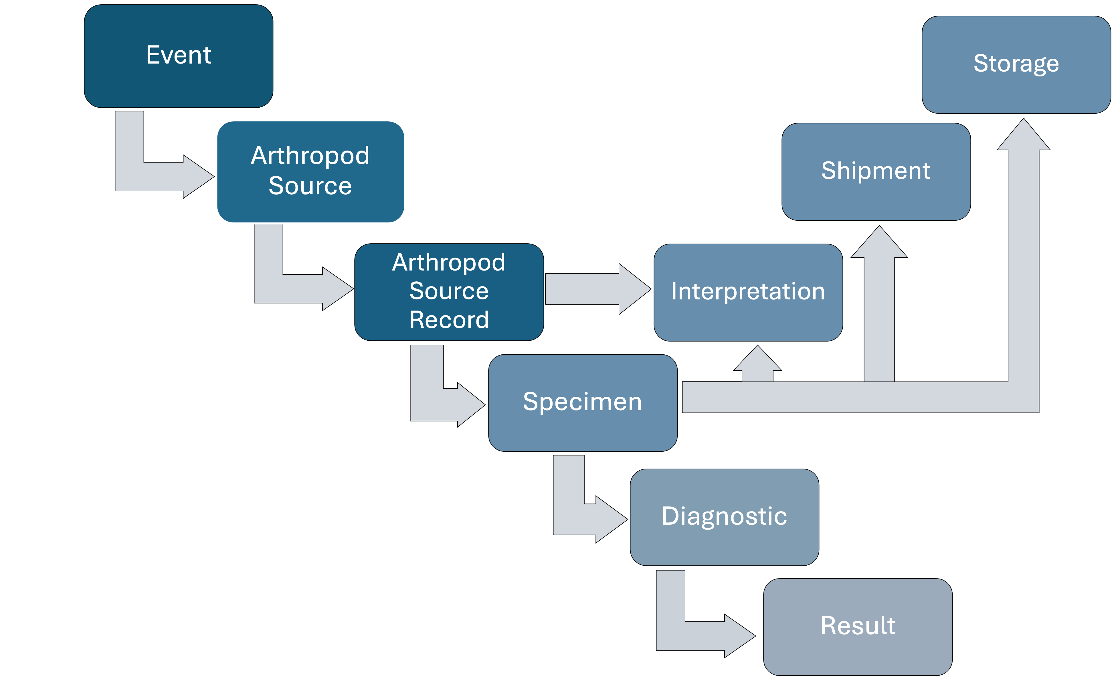
Figure 11: Detailed relationships of Arthropod Source in the data model.
If the interest of the Surveillance Activity is at the individual arthropod level (e.g., butterflies with problems in their wings or with parasites), then the user should consider these arthropods as Animal Sources. Arthropods from Animals Sources (attached ticks, lice, fleas, mites) are Specimens (see “Specimens”) from those Animal Source and not Arthropod Sources.
An Arthropod Source Record represents the set of arthropods collected from an Arthropod Source at time t . For example, mosquitoes captured using a CO₂ trap.
Key attributes of a Arthropod Source Record include:
Other attributes are provided in the Data Dictionary (under construction)
A key characteristic of an Arthropods Source Record is that there can be several species under the Source Record level. For example, mosquitoes collected from a specific site (Source) will be identified and counted by species in the Arthropod Source Record.
When arthropods are collected without effort (No Effort), for example, opportunistically using a brine shrimp net, then the collection method is reported at the Arthropod Source Record level. When the collection implies an effort, the methodology can be inferred from the Point Collection.
An Environmental Source is a site where measurements on environmental properties can be made or samples can be collected from. An Environmental Source can be observed, tested, and provide abiotic or biotic Specimens. An Environmental Source cannot be shipped or stored.
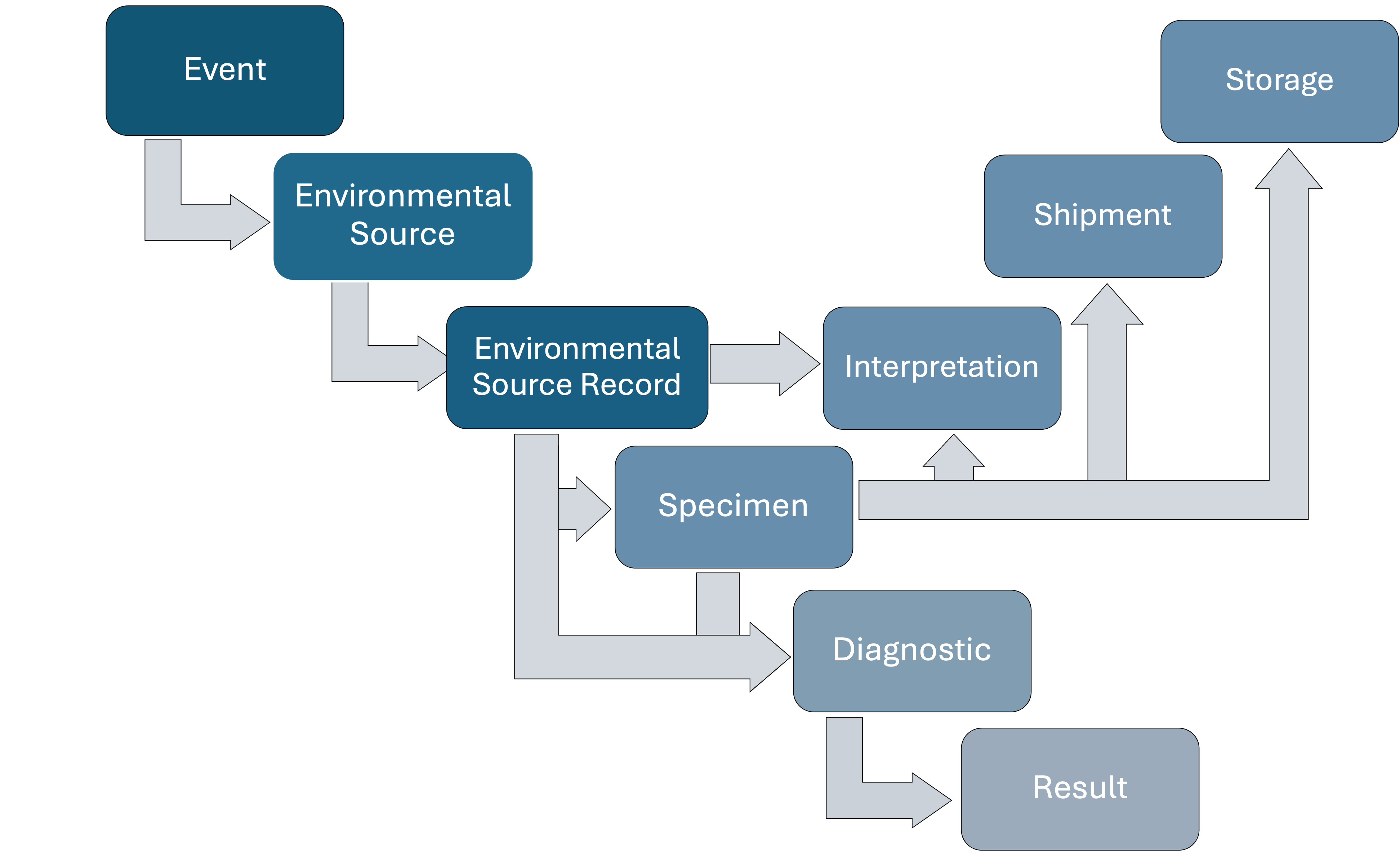 Figure 12: Detailed relationships of Environmental Source in the data model.
An Environmental Source Record is a site where measurements on environmental properties are made or samples are collected at time t. Many types of samples can be obtained from a Environmental Source Record, for example, feces (with unknown animal origin), water, sediment, and air.
Diagnostics can be conducted on site without requiring the retrieval of specimens and, therefore, they apply to the Environmental Source Record directly. For example, measuring the turbidity of the water at the Environmental Source at time t. The type of substratum used for these direct Diagnostics (e.g., water, air, etc) must be provided.
Key attributes of a Environmental Source Record include:
Other attributes are provided in the Data Dictionary (under construction)
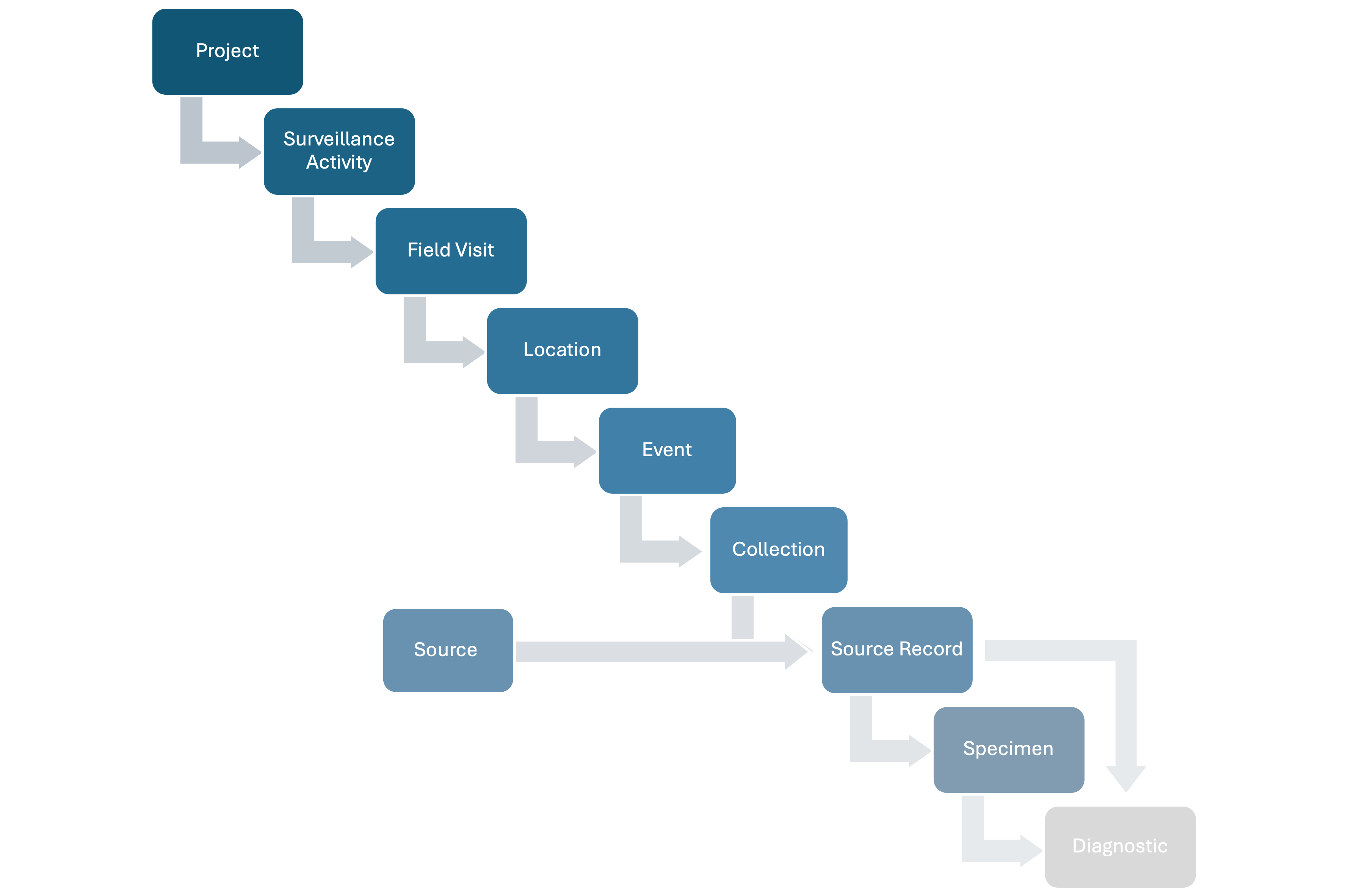 Figure 13: Position of Specimens in the data model.
In the data model, a Specimen refer to a sample collected to conduct Diagnostics (see “Diagnostics”).
Specimens originate from the following:
Sampled Group Source Record (e.g., urine collected under a bat roost and the urine cannot be linked to a specific individual of the roost) and Animal Source Records (e.g., an oral swab from an individual animal). These Specimens may consist of either a single sample type (e.g., blood) or multiple tissue types (e.g., mixed blood and saliva).
Pools of arthropods comprised of the arthropods present at an Arthropod Source Record.
Samples collected from the environment with abiotic origin (e.g., water) or biotic origin (e.g., feces found in the field)
Key attributes of a Specimen include:
Other attributes are provided in the Data Dictionary (under construction)
Specimens can be stored, and their storage may change multiple times as they are transferred within and between facilities. Similarly, Specimens can be shipped multiple times.
Specimens without any amount of tissue left remain in HAWK so the last Storage, Shipment, and use in Diagnostics can be traced.
In situations where space or materials are limited, it is possible that multiple Specimens are stored in a unique container. This approach is clearly not ideal because it can lead to cross-contamination and make actual Specimen tracing more complex. However, the data model has a Container Identifier as a Specimen property (see Data Dictionary). Specific properties for each individual Specimen within the container, such as type, quantity, etc, should allow their visual identification within the container.
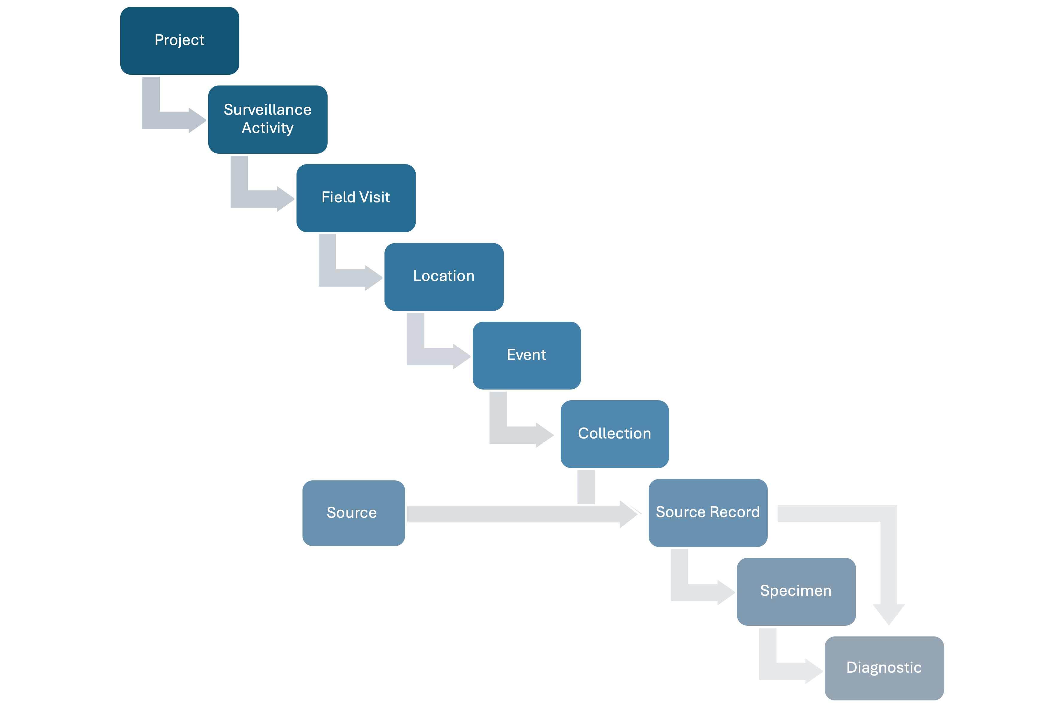 Figure 14: Position of Diagnostic in the data model.
Diagnostics in the data model encompass various methods used to measure health targets of biological, chemical, physical or physiological nature in either Records of Animal, Group, and Environmental Sources or Specimens obtained from Source Records of any type. Diagnostics can range from the clinical examination of an Animal Source to advanced techniques such as metagenomics to sequence nucleic acids present in a Specimen.
Diagnostics report the type and name of the diagnostic method used and who conducted the diagnostic. The actual outcome of a Diagnostic is recorded as a Result (see “Results” below). A single Diagnostic can detect one or multiple targets; therefore, each Diagnostic can have more than one Result.
Key attributes of a Diagnostic include:
Other attributes are provided in the Data Dictionary (under construction)
In the data model, Diagnostics can be conducted in a Laboratory or similar (see Laboratory below) but it is also possible to include field-based assays.
Anomalies in a Group Source Record or Animal Source Record detected through observation or smell (e.g., missing an ear) are not recorded through a Diagnostic but as characteristics of the Group Source Record or Animal Source Record. Similarly, abnormal findings at a specific site detected through observation, smell, or hearing (e.g., smoke) are not recorded through a Diagnostic but they are a property of the Event.
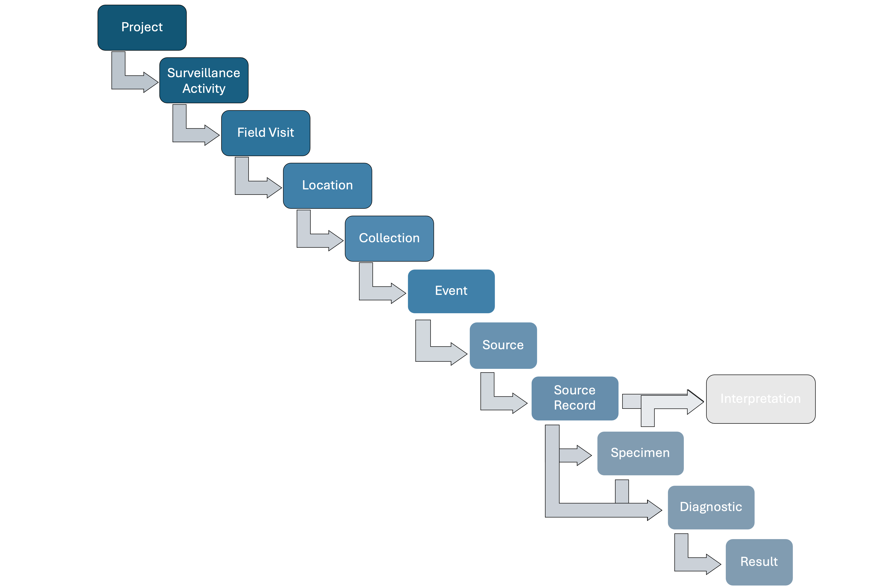 Figure 15: Position of Result in the data model.
A Result request information on the Diagnostic outcome for a single target, in quantitative or qualitative units. A Result can be more specific, reporting the outcome with respect to a gene of a specific pathogen that the Diagnostic can detect. For example, a real-time PCR can target the detection of Influenza A nucleic acid by the amplification of genes X and Z. The Result reports the Ct value for the amplification of either gene X or Z of Influenza A viruses.
Key attributes of a Result include:
Other attributes are provided in the Data Dictionary (under construction)
The Result of a Diagnostic can be interpreted as positive, negative, or undetermined, based on the case definition for the parent diagnostic test (reported in the Surveillance Activity metadata).
A Storage indicates the facility where carcasses of Group Source Records, the carcass of an Animal Source or Specimens are stored. The storage position can be as specific as the exact row of a cryo box in which a vial containing a Specimen is located.
Key attributes of a Storage include:
Other attributes are provided in the Data Dictionary (under construction)
Multiple Storage units can be added for each Source or Specimen. In the case of Specimens, the recorded Storage units allow to track the current amount available versus the original amount and the reason of the differences over time.
A Shipment indicates the movement of carcasses of Group Source Records, the carcass of an Animal Source or Specimens across facilities.
Key attributes of a Shipment include:
Other attributes are provided in the Data Dictionary (under construction)
In the data model, a Specimen can receive an Interpretation with respect to the hazards targeted in the corresponding Surveillance Activity based on a case definition for a positive, negative, or undetermined Specimen provided in the Surveillance Activity metadata. The Interpretation follows the Results from the Diagnostics conducted using the Specimen and their interpretations.
Specimens can receive multiple Interpretations if they are used in several Diagnostics targeting different hazards.
The presence of a single target (e.g., SARS-CoV-2) is assessed in a Specimen using three different Diagnostics. The first and second Diagnostic are positive, whilst the Result for the third Diagnostic is negative. Based on these mixed Results, the SARS-CoV-2 status of the Specimen must be interpreted (positive, negative, inconclusive).
In the data model, a Source Record can receive an Interpretation with respect to the hazards targeted in the corresponding Surveillance Activity based on a case definition for a positive, negative, or undetermined Source Record provided in the Surveillance Activity metadata. An Interpretation for a Source Record depends on the field findings, Interpretations for the Specimens of the corresponding Source Record, Results of Diagnostics conducted with the Source Records (for Group Source Records and Animal Source Records) and Necropsy findings (for Animal Source Records).
A Source Record can receive multiple Interpretations depending on the hazards targeted.
The presence of a single target (e.g., Foot and mouth disease virus) is assessed in an Animal Source Record using three different Diagnostics completed with one Specimen and the results of a Necropsy. The Results for the first and second Diagnostic are positive, whilst the Result for the third Diagnostic is negative. The Interpretation for the Specimen is “posiive”. Moreover, the Necropsy do not reveal any important lesions. Based on these Results, the Foot and mouth disease virus status of the Animal Source Record must be interpreted (positive, negative, inconclusive).
Key attributes of a Interpretation include:
Other attributes are provided in the Data Dictionary (under construction)
The structure of the data model could be enough to record the hierarchical data. For example, rangers collecting information during patrols (Tracks Effort) in Protected Areas (Locations) at specific points (Events) where dead animals (Source Records) are found at any given time t.
However, the data in another Surveillance Activity with animal captures could have the following data structure:
Looking at the examples above, the units Location, Event, and Effort might not be enough to accommodate the data structure of certain designs. To solve this limitation, the data model allows to tag units (from Surveillance Activities up to Diagnosis) with labels that allow to further group them in different categories.
The following example shows a potential data structure for the sampling of animals in markets:
In this case, tagging Events with the corresponding stall id and with the corresponding vendor id will group them by stall and vendor. Locations can be tagged with the corresponding neighborhood and city.
Tags can group units spatially, temporally, or in another way. Tags can be nested, non-nested, or a combination of both.
Unavoidably, the number of Tags, what Tags represent, what they group, the number of categories per group will vary among Surveillance Activities. Therefore, Tag properties must be reported in the Surveillance Activity metadata. In the data model, the only default property for each Tags are the name and the type.
Field Visits, Locations, Events, Sources, Source Records, Specimens, and Diagnostics usually belong to a single Surveillance Activity. This is the Surveillance Activity that lead to generate these data, creating Events, Sources, and Specimens.
However, units can belong to more than one Surveillance Activity:
When a Surveillance Activity uses stored carcasses to test them for cardiomegaly and the carcasses were obtained through a previous Surveillance Activity
When a Surveillance Activity uses stored specimens to test them for a health target an the carcasses were obtained through a previous Surveillance Activity
An agency decides to test stored deer samples for SARS-CoV-2. The blood samples have been stored for over a year. The current surveillance initiative is only recycling the samples already collected, its funding source is different, and its objectives and methods are different
In the data model, a new Surveillance Activity is created documenting the new methods and objectives. Then, the Source Records or Specimens to be reused and their parent Field Visits, Locations, Efforts, and Events are added to this new Surveillance Activity. The exception is a field for Field Visit that request its origin. The Field Activity in the original Surveillance Activity has this field completed with “Current Surveillance Activity” whilst in the new Surveillance Activity it should be completed with “Previous Surveillance Activity”. Moreover, Field Visits with origin as “Previous Surveillance Activity” need to report the original Surveillance Activity ID.
New Necropsy, Specimens, Diagnostic, Result, and Interpretation units can be added under the new Surveillance Activity
As explained in the section “Surveillance Activity”, an outbreak investigation is a Surveillance Activity. An outbreak can be first detected by rangers patrolling a protected area. Therefore, the Field Visit, Location, Event, and Source Records by the rangers belong to the Ranger Patrol” Surveillance Activity and also to the outbreak investigation Surveillance Activity.